
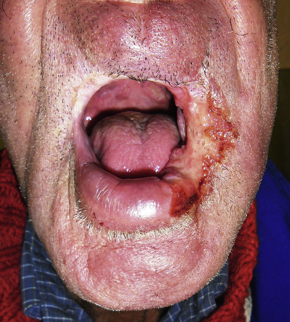
A 65-year-old man, actively engaged in farming activities, with a 90 pack-year smoking history, presented with a painless ulcerated lesion on the left labial commissure. He was referred to our department because of suspected neoplasia. The lesion progressed slowly through a 2-year period. Oral examination revealed an extensive ulcer, although flat and with no signs of orbicularis oris muscle infiltration (Fig. 1), involvement of mandibular division of facial nerve, or suspicious neck lymph nodes metastasis. A representative incisional biopsy was performed. Histopathological examination exhibited chronic granulomatous inflammation on conventional hematoxylin–eosin staining. Grocott-Gomori staining showed fungal structures. Clinical and pathological evidences supported the diagnosis of paracoccidioidomycosis (PCM). Itraconazole was maintained for six months with satisfactory response to proposed therapy.

PCM is a systemic mycosis characterized by acute or chronic tissue inflammation caused by Paracoccidioides brasiliensis, a pathogenic thermally dimorphic fungus that is endemic to Latin America.1 Active agricultural laborers, masons and civil construction workers, or those who have a history of agricultural labor are at greater risk for PCM.1 Men are nine times more likely to be diagnosed than women.1 The cutaneous and mucosal lesions result from hematogenous dissemination, either by contiguity or rarely by direct inoculation under the tissue. Signs and symptoms are related to the affected site, and usually appear as visible lesions or non-specific symptoms.1 In cases of mucocutaneous disease, the oral and nasal cavities are the most commonly affected sites.2
Similarly to PCM, head and neck carcinomas usually present with exophytic, infiltrative or ulcerative lesions, affecting middle-aged and elderly white men, mostly those with smoking and/or drinking habits.3 As risk factors and clinical manifestations are similar to those of head and neck carcinomas, a differential diagnosis is necessary.
Author contributionsFábio Muradás Girardi contributed to conception, design, acquisition of data, analysis and interpretation of data; drafted the article and revised it critically for important intellectual content; gave final approval of the version to be published; and agreed to act as guarantor of the work (ensuring that questions related to any part of the work are appropriately investigated and resolved).
Maria Lúcia Scroferneker contributed to conception, design, and interpretation of data; drafted the article and revised it critically for important intellectual content; gave final approval of the version to be published; and agreed to act as guarantor of the work (ensuring that questions related to any part of the work are appropriately investigated and resolved).
Conflicts of interestThe authors declare no conflicts of interest.
The author would like to thank Dr Bruno Schinke for his assistance with pathological examination.
