

Bacterial vaginosis (BV) is the most common cause of vaginal discharge in women of reproductive age. The purpose of this study was to determine the frequency of BV in Bulgarian pregnant and nonpregnant women from several age ranges and to compare three different laboratory methods for Gardnerella vaginalis detection in patents suffering from BV.
MethodsBetween September 2011 and June 2012, 809 women of 16–40 years of age separated in two major groups: nonpregnant – 469 (355 with and 114 without symptoms) and pregnant – 340 (213 and 127 respectively) were enrolled for the study. The women underwent three different laboratory tests simultaneously: scoring of Gram staining of vaginal smear, culture, and polymerase chain reaction (PCR) assay for G. vaginalis.
ResultsThe microscopic method detected high frequency of BV in symptomatic (57%) whereas only a minority of asymptomatic subjects (14%) were detected. G. vaginalis-associated BV was diagnosed in approximately equal proportions when evaluated with PCR and microscopic method for both pregnant and nonpregnant women. The comparative analysis of microscopic evaluation, culture and PCR assays demonstrated greater concurrence (about 90%) between Gram staining and PCR detection for BV, than both methods compared to culture. The combination of microscopy and PCR turned out to be very reliable and repeatable for detecting G. vaginalis-associated BV.
ConclusionsThis is the first comparative investigation on the epidemiology of G. vaginalis-associated BV in Bulgaria. The established highest frequency in the young Bulgarian women (21–30 years) is alarming and should be considered in prophylaxis and reproductive programmes.
Bacterial vaginosis (BV) is the most common cause of unpleasant vaginal odor and discharge in women of reproductive age.1,2 It is induced by an imbalance in naturally occurring microflora. Any change in the resident flora including reduction of lactobacilli allows for different anaerobic bacteria to gain a foothold and multiply.3–7 Nevertheless the process is multifactorial and the initial mechanism of replacement of normal lactobacillary flora by opportunistic pathogens in vaginal ecosystem and the role of intrinsic host factors still remains unclear, requiring more research to be conducted.5–8 The essential participants in pathological polymicrobial associations, which could be used as markers for BV, are Gardnerella vaginalis (that grows under appropriate microaerophilic conditions) and anaerobic Atopobium vaginae.3–8 Other microorganisms involved in BV microbiota are very diverse and include anaerobes, such as Peptostreptococcus spp., Mobiluncus spp., Prevotella spp., Bacteroides spp., Fusobacterium spp., and facultative anaerobes.6–11 It is not clear yet if the BV is a sexually transmitted disease, but it is more common in promiscuous women with hazardous sexual behavior (with multiple and/or new sexual partners; or with female partners, sex during menses).12–18 BV can be an independent risk factor for acquisition of any other sexually transmitted infection.1,15,17 It has also been shown to be a cause for serious health problems as preterm birth, postpartum fever, development of endometritis, post-hysterectomy or postabortal sepsis, and pelvic inflammatory disease.19–21
The purpose of this study was to determine the frequency of BV and G. vaginalis-associated BV in Bulgarian pregnant and nonpregnant women from different age ranges and to compare three distinct laboratory methods for G. vaginalis detections in patients suffering from BV.
MethodsPatients and clinical samplesFrom September 2011 till June 2012 we obtained vaginal samples from 568 women with evident clinical symptoms of vaginal discharge and from 241 asymptomatic women of reproductive age (range 16–40 years). No women had received antimicrobial therapy for at least a week before examination. According to the pregnancy status, subjects were divided into two groups: nonpregnant – 469 women (355 with and 114 without symptoms), and pregnant – 340 women (213 and 127 respectively).
Gram stainingFrom the vaginal samples we prepared smears and classified them into three major groups, using the Nugent scale (range from 0 to 10)22 and the modified scoring method with five grades of flora described by Ison and Hay.23 The first group was comprised of subjects with normal vaginal flora – NVF (Nugent score 0–3; Ison/Hay score 0-I). The second group – with transition between normal flora and BV – TVF (Nugent score 4–6; Ison/Hay score II), the third group was with BV (Nugent score 7–10; Ison/Hay score III). The latter group was subdivided in two subgroups IIIA (true BV) and IIIB – BV, more rare type that was just outside the used scoring criteria and there were no positive data from other investigations (complicated with other vaginal pathogen – single areas with polymorphonuclear leukocytes and Trichomonas vaginalis or Candida spp.).
The use of Amsel's criteria was based on some clinical symptoms that could not be standardized, so we did not include them in the assessment, but we evaluated the most important and significant laboratory indication for BV which was confirmation that more than 20% from the total cell population were clue cells in the oil immersion fields of the vaginal smear that coincides with Nugent score 7–10 and Ison/Hay score III.22–24
CultureThe samples were cultivated in aerobic conditions on non-selective sheep blood agar and MacConkey agar (for residentl microflora) and Sabouraud's agar for Candida spp.
For detection of G. vaginalis we used Columbia blood agar base with G. vaginalis Selective Supplement SR0119E, Oxoid (with gentamicin and nallidix acid) in microaerophilic atmosphere (5–10% CO2) at 36°C for 48–72h. The Gram-negative or Gram-variable short rods, transparent colonies, β-hemolytic on human blood agar, catalase-negative, Glucose, Prolin, ONPG positive, were presumptively identified as G. vaginalis using Remel RapID NH.
The presence of T. vaginalis in vaginal samples was detected by its morphological characteristic of microscopic strain.
DNA isolationTotal DNA from vaginal samples was isolated using the DNA-sorb-AM nucleic acid extraction kit (AmpliSens) according to the manufacturer's guidelines.
Polymerase chain reaction (PCR) assayA species-specific PCR assay for the detection of G. vaginalis targeting the 16S rRNA gene was performed. The oligonucleotides used as primers for amplification were GV1-F (5′-TTACTGGTGTATCACTGTAAGG-3′) and GV3-R (5′-CCGTCACAGGCTGAACAGT-3′) synthesized by Alpha DNA.25 They were verified for specificity using the BLAST program.
PCR was carried out in a total volume of 25.0(L and the final concentration of the mix for each sample contained: 0.25(M of each primer, 0.2mM dNTPs, 1х Reaction Buffer, 2.0mM MgCl2 and 1.5U Taq DNA polymerase (Prime Taq™ DNA Polymerase, GENET BIO). The DNA was amplified using the following protocol: an initial denaturation (94°C for 5min), followed by 30 cycles of denaturation (94°C for 45s), annealing (60°C for 45s) and extension (72°C for 45s), with a single final extension of 7min at 72°C. PCR products were separated in 1% agarose gel for 45min at 140V, stained with ethidium bromide (0.5μg/mL) and detected by UV transillumination (wavelength 312nm). Amplified genes were identified on the basis of their expected fragment size (331bp).
Statistical analysisThe data were analysed using Chi-square and Fisher's exact tests. The results are expressed with calculated standard deviations (SD). We considered p values of ≤0.05 to indicate statistical significance.
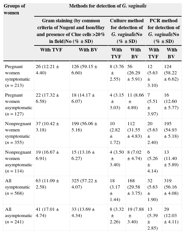
Results and discussionThe data from the three procedures for BV and G. vaginalis detection in different groups of women are presented in Table 1.
Concordance between the groups of women on the basis of three different laboratory methods.
| Groups of women | Methods for detection of G. vaginalis | |||||
|---|---|---|---|---|---|---|
| Gram staining (by common criteria of Nugent and Ison/Hay and presence of Clue cells >20% in field)No (%±SD) | Culture method for detection of G. vaginalisNo (%±SD) | PCR method for detection of G. vaginalisNo (%±SD) | ||||
| With TVF | With BV | With TVF | With BV | With TVF | With BV | |
| Pregnant women symptomatic (n=213) | 26 (12.21±4.40) | 126 (59.15±6.60) | 8 (3.76±2.55) | 56 (26.29±5.91) | 12 (5.63±3.10) | 124 (58.22±6.62) |
| Pregnant women asymptomatic (n=127) | 22 (17.32±6.58) | 18 (14.17±6.07) | 4 (3.15±3.03) | 11 (8.66±4.89) | 7 (5.51±3.97) | 16 (12.60±5.77) |
| Nonpregnant women symptomatic (n=355) | 37 (10.42±3.18) | 199 (56.06±5.16) | 10 (2.82±1.72) | 112 (31.55±4.83) | 20 (5.63±2.40) | 195 (54.93±5.18) |
| Nonpregnant women asymptomatiс (n=114) | 19 (16.67±6.91) | 15 (13.16±6.27) | 4 (3.50±3.40) | 8 (7.02±4.74) | 6 (5.26±4.14) | 13 (11.40±5.89) |
| All symptomatic (n=568) | 63 (11.09±2.58) | 325 (57.22±4.07) | 18 (3.17±1.44) | 168 (29.58±3.75) | 32 (5.63±1.90) | 319 (56.16±4.08) |
| All asymptomatic (n=241) | 41 (17.01±4.74) | 33 (13.69±4.34) | 8 (3.32±2.26) | 19 (7.88±3.40) | 13 (5.39±2.85) | 29 (12.03±4.11) |
SD, standard deviation.
Our results from PCR assay are shown in Fig. 1.
. The product size is 331bp. From right to left: 100bp Ladder and 9 (+) positive samples.")
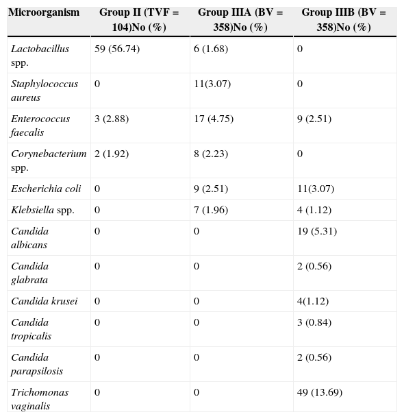
The group distributions of the additive isolates from aerobic cultures are summarized in Table 2.
Isolate distribution among 104 women with diagnosis TVF and 358 women with BV according to Gram staining.a
| Microorganism | Group II (TVF=104)No (%) | Group IIIA (BV=358)No (%) | Group IIIB (BV=358)No (%) |
|---|---|---|---|
| Lactobacillus spp. | 59 (56.74) | 6 (1.68) | 0 |
| Staphylococcus aureus | 0 | 11(3.07) | 0 |
| Enterococcus faecalis | 3 (2.88) | 17 (4.75) | 9 (2.51) |
| Corynebacterium spp. | 2 (1.92) | 8 (2.23) | 0 |
| Escherichia coli | 0 | 9 (2.51) | 11(3.07) |
| Klebsiella spp. | 0 | 7 (1.96) | 4 (1.12) |
| Candida albicans | 0 | 0 | 19 (5.31) |
| Candida glabrata | 0 | 0 | 2 (0.56) |
| Candida krusei | 0 | 0 | 4(1.12) |
| Candida tropicalis | 0 | 0 | 3 (0.84) |
| Candida parapsilosis | 0 | 0 | 2 (0.56) |
| Trichomonas vaginalis | 0 | 0 | 49 (13.69) |
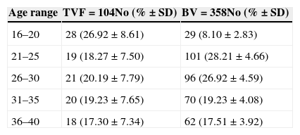
The correlations between some demographic parameters (i.e. age range) with presence of G. vaginalis and BV are demonstrated in Table 3.
Association of age range with BV according Gram staining.
| Age range | TVF=104No (%±SD) | BV=358No (%±SD) |
|---|---|---|
| 16–20 | 28 (26.92±8.61) | 29 (8.10±2.83) |
| 21–25 | 19 (18.27±7.50) | 101 (28.21±4.66) |
| 26–30 | 21 (20.19±7.79) | 96 (26.92±4.59) |
| 31–35 | 20 (19.23±7.65) | 70 (19.23±4.08) |
| 36–40 | 18 (17.30±7.34) | 62 (17.51±3.92) |
SD, standard deviation.
G. vaginalis was most frequently present in samples obtained from Bulgarian women in age range 21–30 years.
For achieving the goal of this investigation, the Nugent's score22 was taken as gold standard and the results from the other methods were compared with this score. As already published by other authors it was preferable to recommend the use of Ison/Hay method,23 because in that way we could evaluate vaginal smears in five more clearly distinguished grades with better segregation of the vaginal microflora.9,26,27 Using Gram straining in this study we detected high frequency of BV in symptomatic subjects (57.22%), in contrast to asymptomatic Bulgarian women (13.69%). We could also prove TVF in 11.09% of symptomatic and in 17.01% of asymptomatic subjects. Our results for TVF frequency are in unison with previously reported data in other studies from France and Australia.3,28 BV frequency in the present study with Bulgarian women is similar to that found in Nigerian women, with a slightly higher rate in our population.29 We have shown that BV occurs in approximately equal proportions when evaluated with the microscopic method for both pregnant and nonpregnant symptomatic and pregnant and nonpregnant asymptomatic women. These results are in contrast to data obtained by other authors, claiming that BV is more frequent during the pregnancy, which we could explain by separating our groups according to the symptoms, and not only by pregnancy status. So far all studies revealed different and sometimes conflicting results for BV epidemiology.1,2,9,10,27,29
We found with PCR that pregnant symptomatic patients with BV and TVF were positive for G. vaginalis in 58.22% (very similar to the 59.15% with Gram straining) and 5.63%, respectively. The group of nonpregnant symptomatic women had positive samples in 54.93% (56.06% using Gram straining) for BV and 5.63% for TVF (Table 1).
Using a selective agar media the isolation rate of G. vaginalis as marker for BV was significantly lower – only in half of the cases (Table 1). The comparative analysis of microscopic evaluation, culture and PCR assays demonstrated greater concurrence (about 90%) between Gram staining and PCR detection for BV, than both methods compared to culture (Table 1). We found that 89% of BV and only 28% of TVF (where clue cells were significantly less than in BV) groups, according to the microscopic criteria, had positive PCR for G. vaginalis. All PCR positive results for G. vaginalis had either BV or T. vaginalis. The combination of Gram staining and PCR methods showed very reliable and repeatable detection of BV, unlike culture, where only about 50% of PCR positive samples had evident growth on the selective agar media for G. vaginalis. PCR assay is the most sensitive method for routing out G. vaginalis (p<0.05), but combination of this test with Gram staining for full characterization in the patient is needed. Gram staining is an easy, fast and affordable method that could be used, especially in low-income countries, instead of PCR, when for various reasons molecular detection is not possible, since the results of both techniques are very similar. The high frequency of G. vaginalis detected by PCR was evident such that this pathogen had a very important role in the aetiology of BV. The results of this study supported the data from previously reported studies where 68–100% of the patients with BV were positive to G. vaginalis.3,9,27
The microbial growth on a non-selective agar media gave useful information for the presence or absence of additional microflora in the pathological process and this procedure should not be skipped, despite having low sensitivity for diagnosing BV. By using this routine method we identified that the most frequently isolated microorganisms colonizing the vaginal mucosa and associated with BV, other than G. vaginalis, were Enterococcus faecalis, Staphylococcus aureus, Corynebacterium spp., Escherichia coli and Klebsiella spp. (Table 2). Their role was unclear, as they might be of transient presence or as they were detected with moderate frequency they could relate to BV aetiology or just be a co-infection. We could not find any confirmed results or evidence for their role published elsewhere. Our data showing Lactobacillus spp. as the predominant bacterial genus present in the vaginal microbiota in the smears and isolates of I-st (NVF) and II-nd group (TVF) according to microscopic evaluation and culture is in line with previously published studies.7,8,24 The Gram-positives were the predominant bacterial microflora in the IIIA group in contrast to the group IIIB where prevailed some Gram-negatives species (Table 2). Some of the isolates such as Candida spp. were detected only among patients of group IIIB, more often in pregnant women and as initial colonization after BV (Table 2). In 13.69% of Bulgarian women T. vaginalis was detected in association with BV (IIIB group). Similar to other reports, trichomoniasis was a frequent infection, and has to be timely diagnosed for its importance as a causative agent of sexually transmitted diseases with difficult and sometimes poor therapeutic response.18,29
BV in Bulgarian pregnant and nonpregnant women was predominantly diagnosed in the age range of 21–25 years (28.21% of all positive samples) and similarly but to a slight lesser extent in the age group of 26–30 years (26.82%). Women in the age ranges 31–35 and 36–40 years had similar detection rates in both groups, which were significantly lower as compared with the previous two age groups. In the youngest group (age below 20 years) BV was detected in only about 8%, whereas TVF was found in 26.92% of women in this age group. Our finding did not differ significantly from those published for Indian women, where BV prevalence in age group of 26–30 years was 23% and in 7% among the youngest group (15–20 years).30 The only difference between the Indian results and ours was in the group of 21–25 years, where BV was more frequent among Bulgarian women.
ConclusionTo our knowledge, this is the first comparative study utilizing three different laboratory methods that focuses on the epidemiology of G. vaginalis-associated BV in Bulgaria. Although PCR is the most sensitive method for the detection of G. vaginalis, but for the full characterization of the smears the joint application of PCR and Gram staining is the best choice. An important note is that Gram staining results are compatible with PCR results, since this method is fast, easy and inexpensive, so that it could be used in developing countries, where and when molecular techniques are not available.
The high frequency in Bulgarian young women found in this study is alarming, since BV increases woman's susceptibility to HIV, HPV and other important sexually transmitted diseases. Therefore BV has to be correctly and timely diagnosed in order to be adequately treated.
Further investigations regarding other pathogens involved in BV such as A. vaginae and Mobiluncus spp. are warranted.
Conflict of interestAll authors declare to have no conflict of interest.
The authors would like to thank Dr. MIkana Shtereva (Medical center Nadejda), Dr. K. Tosheva (Medical center Pentagram), Dr. A. Kosturska and Dr. M. Ilieva (Medical center VIP Clinic), Dr C. Jordanova (Individual medical practice) for providing vaginal samples and demographic data of the patients included in this study.
