


A 7-year-old Mexican male presented to a hospital in Monterrey, Mexico with a 1-week history of persistent abdominal discomfort and loose stools. One month before presentation, the patient had traveled on vacation to Cuba and recalled eating ceviche (a popular raw fish dish in Latin America). He denied vomiting, anorexia, fever or weight loss, and had no other significant medical history. Physical exam was unremarkable. Routine blood and biochemical tests were normal.
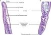
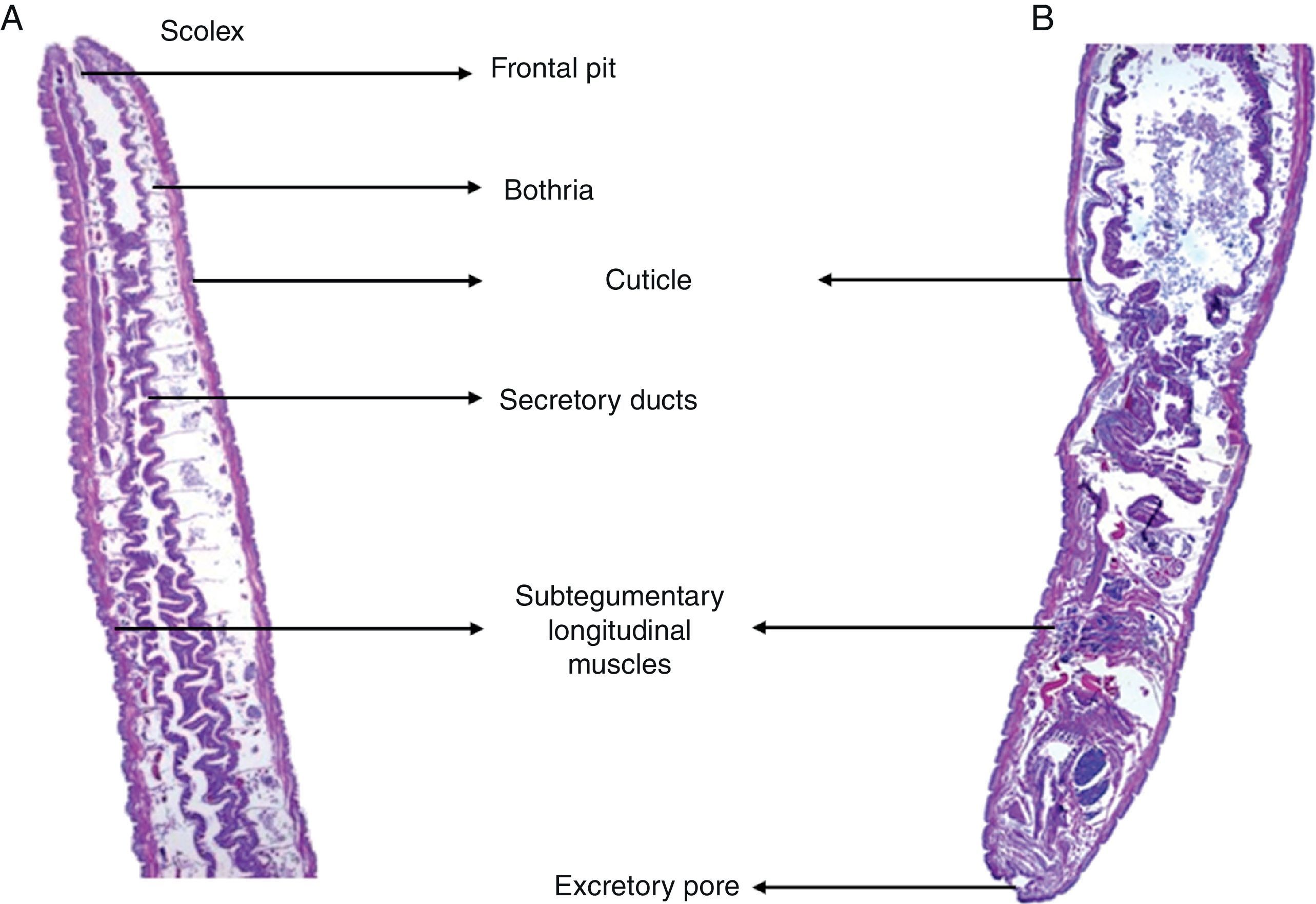
An upper endoscopy detected a larva attached to the duodenal wall. After endoscopic extraction of the worm, symptoms were relieved. Further examination of the larva allowed identification of a plerocercoid larva of Diphyllobothrium sp. (Fig. 1) based on the morphological and histological features described in previous publications.1,2 The larva measured 2.8cm in length. The epithelial morphology of the dorso-ventrally flattened larva's body was typical, with apparent segmentation, traces of the bothria in the anterior end, and invagination at the posterior end. The mid-body presented a single poorly developed layer of subtegumentary longitudinal musculature. Diagnosis of the worm at the species level was not achieved because the adult stage was needed and molecular confirmation was not possible.
Here, we report the first diphyllobothriosis case in Mexico, which was imported from Cuba. Diphyllobothriosis is a widespread fish-borne zoonosis acquired by eating raw or undercooked fish contaminated with plerocercoid larvae of the tapeworm Diphyllobothrium.3,4 Common raw or poorly cooked fish dishes include sushi, sashimi, ceviche, carpaccio, and others. The disease is endemic in the Arctic regions and several parts of Europe, Asia, and the Americas. Given that the disease is usually caused by the adult form of Diphyllobothrium spp., we consider this to be an unusual case presentation in which the larva, attached with its scolex to the intestinal epithelium, was found before becoming an adult.
Author contributionsElba G. Rodríguez-Pérez took part in conception of the manuscript and data acquisition. Kevin Escandón-Vargas drafted the manuscript and reviewed the literature. Jenniffer A. Castellanos contributed to the analysis and interpretation of data.
Conflicts of interestThe authors declare no conflicts of interest.
