


The pathogenesis of cutaneous and mucosal leishmaniasis is associated with different immune responses. Vitamin D may modulate the immune system. Here we evaluate the association of vitamin D levels with the severity of the clinical forms of cutaneous and mucosal leishmaniasis.
MethodsWe conducted an observational study evaluating the association between vitamin D levels, disease severity and therapeutic response in patients with cutaneous and mucosal leishmaniasis. Additionally, we conducted a cross-sectional study to compare vitamin D levels in patients with leishmaniasis and healthy subjects. Hypovitaminosis D was defined as a serum level of 25 (OH) D < 30 ng/mL.
ResultsIn patients with leishmaniasis, vitamin D serum levels were 38.5 ± 11.54 ng/mL, and 37.5 ± 10.43 ng/mL in healthy subjects The prevalence of hypovitaminosis D was 23.3% and 20.0%, respectively (p = 0.72). There was no correlation between vitamin D serum levels, disease severity, and healing time in the mucosal leishmaniasis group.
ConclusionVitamin D levels are not associated with neither susceptibility nor severity of tegumentary leishmaniasis.
Cutaneous leishmaniasis (CL) and mucosal leishmaniasis (ML) are clinical forms associated with Leishmania (Viannia) braziliensis infection with high prevalence in South and Central America according to the World Health Organization. The parasite is transmitted to humans by Lutzomyia phlebotomies biting. After skin penetration, the lesion starts as papules, and later, the classical well-limited ulcer with raised edges becomes evident. After the infection, although a strong Th1 immune response inhibits parasite multiplication, leishmaniasis persists and the exaggerated inflammatory response causes pathology. 1,2 (ANTONELLI, 2005, CARDOSO, 2015).
About 3% of CL patients develop a more aggressive disease in their mucosa. In ML, patients develop ulcers and crusts mainly on the nasal mucosa that can evolve to destructive forms of the septum and other nasal support structures.3 (LESSA, 2012). Pathophysiology of different clinical forms can be explained by parasite-host interaction, immune response presentation, and genotypic difference among L. braziliensis.
Vitamin D is a fat-soluble hormone whose classic action is related to calcium metabolism and maintenance of bone health. About 80% of its acquisition comes from the endogenous conversion in the skin. The UV-B rays irradiation transforms the skin precursor 7-dehydrocholesterol into cholecalciferol. The other 20% is acquired through the diet. Ergosterol and cholecalciferol, respectively vitamin D2 and D3, are the two nutritional forms of vitamin D. After being transported to the liver and kidney where it undergoes hydroxylation and vitamin D takes its active form: 1.25 (OH)2 D. The connection between vitamin D and its receptor VDR induces cell differentiation, apoptosis, adhesion, inflammation, and modulates the immune system. In the innate immune response, vitamin D induces the gene transcription of cathelicidin13(WANG, 2004), an essential antimicrobial peptide in the barrier organs. In the adaptive immune response, vitamin D directly affects the differentiation of CD4+ T lymphocytes by inhibiting Th1 (IFN-γ production) and stimulating Th2 cell development (IL-4, IL-5, and IL-10 production) in BALB/c and C57BL/6 mice.4 (BOONSTRA, 2001).
Previous studies correlating vitamin D and leishmaniasis were restricted to in vivo work. Ehrchen et al., 2007 observed that C57BL/6 knock-out for vitamin D receptor (VDR-KO) mice was more resistant to infection when infected by Leishmania major than wild-type mice8This result demonstrates the vitamin D inhibitory influence on NO production. Whitcomb et al., 201217, found similar results: C57BL/6 VDR KO mice were also remarkably resistant to Leishmania major infection. In addition, VDR-KO mice developed smaller and less parasitized lesions compared to wild mice. Ramos-Martínez et al., 201310, studying BALB/c mice infected with Leishmania mexicana treated with 1.25 (OH) 2D3 observed a dramatic reduction in the size of the lesions compared to the infected and untreated group, indicating better infection control. Bezerra et. al., 20191, evaluated the influence of vitamin D on experimental infection of C57BL/6 and BALB/c mice against Leishmania amazonensis. The animals were submitted to a vitamin D dietary restriction 45 days preceding the infection. Both strains of mice showed smaller lesions when compared to their wild versions; there was no difference in the production of IL-4 and IL-17. However, animals with restricted vitamin D intake had reduced IL-10 levels, indicating that vitamin D contributes to the sensitivity to cutaneous leishmaniasis. Also, it showed that Th1 cell population may be related to the resistance of vitamin D-deficient mice to L. amazonensis.
There was a lack of studies evaluating vitamin D serum levels in patients with tegumentary leishmaniasis due to Leishmania braziliensis in humans. To fill this gap, we proposed to assess if vitamin D deficiency was associated with the severity of L. braziliensis infection and therapy response.
MethodsThis observational study included 30 CL and 30 ML patients recruited in Leishmania Reference Center of Corte de Pedra, Presidente Tancredo Neves, Bahia. The diagnosis was based in a typical cutaneous or mucosal leishmaniasis lesion associated with one of the following criteria: 1) amplification of L. braziliensis DNA by PCR technique in biopsy fragment; 2) growth of L. braziliensis in culture; or 3) finding an amastigote in the anatomopathological examination of the biopsied tissue. We excluded from the study: 1) patients with disseminated leishmaniasis; 2) individuals using vitamin D, multivitamin supplements, anti-convulsants, antiretroviral therapy, oral corticosteroids, rifampicin, cholestyramine, and orlistat users; 3) individuals with chronic kidney failure, disabsorptive intestinal diseases, undergoing bariatric surgery, and pregnant women.
Patients with cutaneous and mucosal leishmaniasis were matched on sex and age ± 5 years old. Healthy control individuals were matched with the mucosal group at age ± 5 years old. The study subjects had their blood collected. Serum was obtained after centrifugation and stored at -70º C until use. Measuring of 25-hydroxyvitamin D levels was performed by chemiluminescence method (Abbott Kit reference 5P02-35, lot 77113UI01, São Paulo- SP). Hypovitaminosis D was considered when serum levels were lower than 30 ng/mL. 25-hydroxyvitamin D serum levels between 30 to 59 ng/mL were considered normal range. Demographic and clinical data such as age, sex, weight, height, illness duration before diagnosis, size of the lesion, and intradermal Montenegro reaction were collected. All subjects had Fitzpatrick's skin phototypes IV, V, and VI.
Written informed consent to participate in the study was obtained from all included patients.
ML patients were staged from I to V following Lessa et al., 2012 criteria (LESSA, 2012)3. Stage I is characterized by nodular lesions of the mucosa without ulcerations; stage II by superficial mucosal ulcerations with concomitant fine granular lesions; stage III by deep mucosal ulcerations with granular tissue formation; in stage IV there is an irreversible perforation of the cartilaginous nasal septum; in stage V the nasal pyramid is impaired because of severe tissue destruction. The mild forms of mucosal leishmaniasis are stages I and II, while the III-V are considered severe (Fig. 1).
 Stages I and II: mild forms of mucosal leishmaniasis. (1C, 1D and 1E) Stages III, IV and V respectively: severe forms of mucosal leishmaniasis. IC, inferior conchae; S, septum.3.")
Clinical aspects of mucosal leishmaniasis at different stages of the disease. (1A and 1B) Stages I and II: mild forms of mucosal leishmaniasis. (1C, 1D and 1E) Stages III, IV and V respectively: severe forms of mucosal leishmaniasis. IC, inferior conchae; S, septum.3.
Patients with leishmaniasis were treated with Glucantime® (Sanofi-Aventis) at a dose of 20 mg/kg/day, with a maximum daily dose of 1,215 mg, Sbv for 20 days in CL and for 30 days in mucosal leishmaniasis group. Intravenous liposomal amphotericin B was the first-choice treatment in elderly patients at a dose of 2 to 3 mg/kg/day, until reaching a total dose of 35 to 40 mg/kg. The patients were followed every 30 days up to 90 days. Cure of CL was defined as complete healing of the lesion and re-epithelization of the tissue without raised borders. The presence of an active lesion characterized therapy failure. In case of failure to antimony therapy, patients received a second course of the drug for another 30-day duration, per the criteria defined by the Brazilian Health Ministry12.
Sample size was calculated using the tool www.openepi.com. Hypothetical hypovitaminosis D frequency in the leishmaniasis population was estimated at 65% (Rolim et al., 2016)11 and hypovitaminosis D was assumed to be two-fold higher in the leishmaniasis group compared to the healthy group. These assumptions resulted in a sample size of 30 patients per group (mucosal leishmaniasis, cutaneous leishmaniasis, and healthy subjects).
Student's t-test was used to compare the serum vitamin D levels between the leishmaniasis group and the healthy group. Ordinal logistic regression was used to assess serum vitamin D levels with therapeutic response in patients with leishmaniasis. Pearson's correlation was employed to assess the correlation between serum vitamin D levels with disease severity in the mucosal leishmaniasis group. A p-value < 0.05 was adopted to assess statistical significance and test hypotheses. The analyses were performed using the IBM SPSS Statistics 20.0 for Windows statistical package. This research was approved by the Research Ethics Committee of the School of Medicine—Federal University of Bahia—Brazil, under the number 2.949.372.
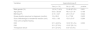
ResultsDemographic features and vitamin D levels in patients with leishmaniasis and in healthy controls are shown in Table 1. While age was similar in the two groups, there was a male predominance among patients with leishmaniasis. There was no difference in vitamin D levels among the groups nor in hypovitaminosis D prevalence.
In the leishmaniasis groups, vitamin D serum levels found were 37.5 +- 11.07 ng/mL in ML patients (n = 30) and 39.5 +-12.09 ng/mL in CL patients (n = 30).
Univariate analysis was performed to test for the association between hypovitaminosis D and clinical data and Montenegro leishmanial skin test (Table 1). However, there was no association of vitamin D levels with sex, age, body mass index (BMI), time until leishmaniasis diagnosis, size of Montenegro's intradermal reaction and therapeutic response (Table 2). Twelve out of the 60 patients with leishmaniasis were lost-to-follow-up after starting treatment; therefore, it was only possible to evaluate therapeutic response of 48 individuals.
Demographic and clinic variables of the patients according to the presence of hypovitaminose D.
A Student t-test also evaluated if there was a statistically significant difference between the serum levels of vitamin D in the presentation of mild mucous leishmaniasis (stages I and II) and severe forms (stages III, IV and V). Fourteen patients with the mild form of the disease had a mean serum vitamin D of 38.20 ± 12.77 ng/mL and while the 15 patients with a severe form of the disease had a mean of 37.40 ± 9.88 ng/ml (p = 0.85). In addition, no association between hypovitaminosis D and therapeutic response was found, with an odds ratio of 1.28 (95% confidence interval: 0.32 to 4.86).
It is worth mentioning that one patient with mucosal leishmaniasis had only a pharyngeal lesion. For that reason, his mucosal leishmaniasis was not staged since the classification is based on nasal mucosa disease.
DiscussionThere were no prior studies on the prevalence of hypovitaminosis D in patients with tegumentary leishmaniasis in an endemic area of Brazil. Herein, we show that vitamin D serum levels were similar in patients with tegumentary leishmaniasis and in healthy subjects. Also, vitamin D serum levels were not associated with tegumentary leishmaniasis severity or therapeutic response.
A 23.3% prevalence of hypovitaminosis D in this study was found in patients with tegumentary leishmaniasis. However, specific characteristics of our sample may have contributed to the low prevalence of hypovitaminosis D. This study was conducted with a rural population involved in professional activities such as farming and fishing, characterized by extensive exposure to solar radiation. This finding is in line with other studies conducted in Northeast Brazil, for example, Monteiro Júnior et al., 20189 found a 4.86% prevalence of hypovitaminosis D in 382 small farmers with a mean age of 57.79 ±15.3 years and a mean serum 25(OH)D levels of 50.4 ± 13.5 ng/mL. This study was performed in Alcântara city, Maranhão state, and hypovitaminosis D was defined as serum 25(OH)D levels < 30 ng/mL
Despite the high variability of vitamin D serum levels due to diverse countries' characteristics, studies worldwide, including in tropical countries, show hypovitaminosis D affecting more than half of individuals.5 (MANDARINO, 2015).
We expected to find a difference between patients with tegumentary leishmaniasis and healthy individuals since vitamin D can influence T cells immunomodulation.6 (LEMIRE, 1984). By its receptor VDR, vitamin D suppresses T helper (Th) lymphocytes proliferation and influences the modulation of cytokines production.6 (LEMIRE, 1984).
In vitro, vitamin D inhibits Th1 response and promotes Th2 cytokines.7 (LEMIRE, 1995). This finding is reinforced by Whitcomb et. al., 201214 experiments in which C57BL/6 VDR KO mice infected with Leishmania major developed smaller and less parasitized lesions and achieved faster healing in comparison to their wild type. In this same study, BALB/c VDR KO mice, known to be unable to mount a Th1 response against Leishmania infection, had not altered susceptibility to L. major nor developed smaller lesions than their wild counterparts. Whitcomb's research demonstrates that VDR suppression or its ligand 1.25-hydroxyvitamin D3 elimination can increase resistance to infection by L. major only in predisposed Th1 responses hosts, indicating an immunomodulatory vitamin D influence on the adaptive immune response.
There are some limitations in our study. First, there is an unbalance in sex between patients with leishmaniasis and the healthy group, likely due to the differential healthy seek behavior between men and women. Second, the study had a small sample size, and we could not access the therapeutic response of 12 patients. Third, no data was available about dietary habits.
ConclusionsVitamin D serum levels do not correlate with the expression or the severity of American tegumentary leishmaniasis.
We want to thank all Corte de Pedra’s patients and participants who took part in this study. Also, we are grateful for the support by the Brazilian Ministry of Science, Technology, and Innovation; the Brazilian Research Council (CNPq) and the Fundação de Amparo à Pesquisa do Estado da Bahia (FAPESB).

- Home
- All contents
- Publish your article
- About the journal
- Metrics