
Respiratory viruses are a possible etiology of infections in pediatric febrile neutropenic patients, considering that they are the most frequent cause of fever in otherwise healthy children.1 We conducted a prospective, observational study, from March to December 2012, in Santa Casa de São Paulo, a busy quaternary public hospital in the largest and most populous city in Brazil, with the objective of evaluating the frequency of respiratory viruses in upper airway secretion samples from febrile neutropenic pediatric patients.
Fever and neutropenia were defined as previously described.2 Febrile neutropenia episodes in the same patient were considered different episodes if they were at least 15 days apart and complete resolution of the previous episode had occurred. Patients were invited to participate in the study within 72h after admission. If agreed, a written consent form was signed by one of the parents or the responsible adult for the patient. After consent, information regarding patient's demographics, clinical and laboratory data was collected. A nasal swab, an oropharyngeal swab and also a saliva sample (collected with Salivette®) were obtained. Viral detection was performed using the FTD Respiratory 21-Multiplex (Fast-Track Diagnostics, Luxembourg), a real-time polymerase chain reaction (PCR) kit capable of detecting 21 agents, including influenza A, H1N1 and B; coronaviruses N63, OC43, 229E and HKU1; parainfluenza 1, 2, 3 and 4; rhinovirus; respiratory syncytial viruses A and B; adenovirus; enterovirus; parechovirus; bocavirus; metapneumoviruses A and B; and Mycoplasma pneumoniae.
Twenty patients were admitted to the hospital during the study, with 38 febrile neutropenia episodes. Five of these patients refused to participate in the study. Of the remaining 15 patients (23 febrile neutropenia episodes), 40% were male, with a mean age of 8.9 years. The most common oncologic diagnosis was acute lymphocytic leukemia, in 10 patients. Other oncologic diagnosis included acute myeloid leukemia, Hodgkin's lymphoma, osteosarcoma, Ewing sarcoma and hepatoblastoma. In 18 of the 23 episodes, a clinical diagnosis of infection was possible, including upper respiratory infection (5) oral candidiasis (5), gastrointestinal infection (3), catheter-related infection (2), lower respiratory tract infection (1), herpes zoster (1), and necrotizing fasciitis (1). Patients had positive blood cultures in 8 of the 23 episodes, and the most common agents were Gram-negative bacteria. Urine cultures were positive in two episodes, both for Gram-negative agents. In eight patients at least one respiratory virus was detected. The most frequent was coronavirus, including 229E, NL63 and OC43 types, present in seven patients. These coronaviruses were found from June to August, which is winter season in our hemisphere. We also detected one rhinovirus, one SRV and one parainfluenza.2 There was viral co-detection in four patients, always including a coronavirus (Table 1). Seven of those eight patients had another febrile neutropenic episode two to five months later. None of them had respiratory viruses detected again. Interestingly, only in three of the eight patients a clinical diagnosis of respiratory tract infection (one with otitis media, one with pneumonia and one with common cold) was present. In three of eight patients bacteria were isolated from a sterile site, but none were related to respiratory disease. None of the patients needed intensive care, mechanical ventilation or vasoactive drugs during hospitalization. There were no deaths.
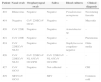
Pediatric neutopenic febrile patients with positive results for respiratory viruses.
| Patient | Nasal swab | Oropharyngeal swab | Saliva | Blood cultures | Clinical diagnosis |
|---|---|---|---|---|---|
| #10 | Rhinovirus | Negative | Negative | Pseudomonas aeruginosa | Necrotizing fasceitis |
| #19 | Negative | CoV 229ECoV OC43 | Negative | – | Mucositis |
| #20 | CoV 229E | Negative | Negative | Acinetobacter sp. | – |
| #21 | CoV 229E | Negative | Negative | – | Pneumonia |
| #24 | CoV 229ECoV NL63 | Negative | Negative | Staphylococcus coagulase-negative | Otitis media |
| #26 | CoV 229ECoV NL63 | CoV 229ECoV NL 63CoV OC43PI2 | CoV 229ECoV NL 63CoV OC43PI2 | – | – |
| #27 | CoV OC43 | Negative | Not collected | – | CRI |
| #29 | SRVCoV OC43 | SRV | – | Common cold |
CoV, coronavirus; PI2, parinfluenzae 2; SRV, syncicial respiratory virus; CRI, catheter related infection.
Although the number of patients and episodes included in our study was low, the results were unique regarding the high frequency of coronaviruses. Previous studies in similar populations have shown that coronaviruses are relatively rare, accounting for less than 5% of all detected respiratory viruses.2,3 One possible reason for our unexpected findings could be the accuracy of coronavirus detection by the RT-PCR method that was used. Some authors have found significant heterogeneity in the accuracy of different multiplex RT-PCR methods for the detection of specific viruses.4 In addition, there is the possibility that not only coronaviruses, but also other respiratory viruses could be detected in asymptomatic individuals. These viruses could possibly be the remaining of a recent infection or could even be part, in some cases and intermittently, of the human virome.5 Larger studies are necessary to evaluate more properly the frequency of coronaviruses in the upper respiratory tract not only in oncologic patients but also in the general population, including asymptomatic individuals. The exact clinical significance of these findings remains to be determined.
Conflicts of interestThe authors declare no conflicts of interest.
