
We hereby describe the clinical and epidemiological features and, outcomes of nine patients with Elizabethkingia meningoseptica infections in two hospitals over a 2-year period. All infections caused by this pathogen were nosocomial, or healthcare associated infections, in hemodialysis settings whereas none was correlated with hospital outbreaks.
We hereby describe the clinical and epidemiological features and, outcomes of nine patients with Elizabethkingia meningoseptica infections in two hospitals over a 2-year period. All infections caused by this pathogen were nosocomial, or healthcare associated infections, in hemodialysis settings whereas none was associated with hospital outbreaks.
An increase of uncommon Gram-negative bacilli has occurred in the last decade in the nosocomial environment. A previous study in Taiwan reported an increased incidence of bacteremia caused by E. meningoseptica between 1999 and 2006, with an incidence rate ranging from 7.5 to 35.6 per 100.000 admissions.1E. meningoseptica is resistant to multiple antibiotics and has been previously described as a pathogen of neonatal meningitis and sepsis,2 as well as a cause of infection among immunocompromised patients.3 It causes significant morbidity and mortality rates may be as high as 50%.2
Infections caused by E. meningoseptica have rarely been identified, but in the last two years we found that several patients were diagnosed with nosocomial infections caused by this agent. The purpose of this study was to describe the clinical and epidemiological features and the outcomes of nosocomial infections caused by E. meningoseptica isolated at two hospitals involving different specialties.
We conducted a retrospective chart review of patients with nosocomial infections caused by E. meningoseptica (CDC),4 from August 2010 to April 2012. Patients diagnosed with E. meningoseptica infections were initially identified from computer databases of the hospital infection committees of the Dante Pazzanese Institute and the Hospital Brigadeiro (São Paulo, Brazil). These two centers are tertiary teaching hospitals containing a total of 356-beds (73 ICU beds) and 150-beds (18 ICU beds), respectively. The former provides care for clinical and surgery cardiac conditions, and the latter for transplant and hematological diseases. The Institutional Ethics Committee approved the study and waived the need for informed consent.
For every patient information was obtained concerning age, weight, gender, hospital course of treatment, antibiotic therapy within 30 days of the E. meningoseptica isolation, time elapsed from hospital admission to the E. meningoseptica infection, length of hospitalization, length of intensive care unit stay (LOS ICU), use of invasive procedures such as peripheral or central catheters, urinary catheter and mechanical ventilation within 30 days of the E. meningoseptica isolates, surgical procedures, antibiotic therapy following infection, time elapsed from the E. meningoseptica infection to death and presence of infection before, during and after isolating E. meningoseptica.
The isolates had been previously identified at the microbiology laboratory of each hospital using the Vitek® system (bioMérieux, France). Four E. meningoseptica isolates obtained from blood cultures were sent to the Adolfo Lutz Institute, a public health reference laboratory, to confirm the identification and to perform susceptibility tests. Classical phenotypic methods and API 20 NE System (Biomerieux, Mercy l’Etoile, France) were used for bacterial identification. The minimum inhibitory concentrations (MICs) were determined by E-test strips (AB Biodisk, Solna, Sweden) for the following antimicrobial drugs: quinolones (ciprofloxacin and levofloxacin), trimethoprim-sulfamethoxazole, minocycline, and vancomycin. These tests were performed according to the recommendations of the Clinical and Laboratory Standards Institute.5 Breakpoints for Staphylococcus aureus were used to interpret the MIC for vancomycin.
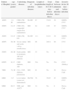
Nine patients were identified, six from the Dante Pazzanese Institute and three from the Brigadeiro Hospital. The clinical characteristics of each patient are presented in Table 1.
Clinical and outcome description of Elizabethkingia meningoseptica infections.
| Patient n°/Hospital | Age (years), gender | Underlying diseases | Diagnosis of infection diseases | Length of hospitalization (days) | Total length of ICU/ICU stay before infection (days) | Time between admission and infection (days) | Invasive device 30 days before infection |
|---|---|---|---|---|---|---|---|
| 1/DPI | 1.4; M | CHD, PH, malnutrition | Pn-MV | 87 | 75/24 | 36 | EI/MV, CVC |
| 2/DPI | 0.4; F | CHD, DS, congenital megacolon, malnutrition | BSI | 84 | 10/9 | 22 | EI/MV, CVC |
| 3/DPI | 0.4; F | CHD, PH, DS | Pn-MV | 111 | 41/21 | 30 | EI/MV, CVC |
| 4/DPI | 68; M | Severe mitral and aortic valves insufficiency | BSI | 66 | 20/14 | 60 | CVC |
| 5/DPI | 64; M | Endocarditis in treatment, AH and diabetes | BSI | 35 | No | 11 | PVC |
| 6/DPI | 81; F | CRF, diabetes, AH | BSI | NA | No | NA | CVC (Shilley) |
| 7/BH | 74; M | CLL, COP, diabetes, pneumonia | BSI | 81 | No | 58 | Port-a-Cath |
| 8/BH | 3; F | Liver failure (cause not determined) | Pn/VM | 76 | 76/45 | 45 | EI/MV, CVC |
| 9/BH | 60, F | NHL and ASCT | BSI | 14 | No | 9 | Port-a-Cath |
| Patient n°/Hospital | Surgery or procedure and length time before infection (days) | Previous exposure to antibiotics 30 days before | Susceptibility tests MICs (μg/mL) | Therapy, time of administration (days) | Associated infections before E. meningoseptica infection | Outcome after infection E. meningoseptica and length between infection and death |
|---|---|---|---|---|---|---|
| 1/DPI | Cardiac catheterization, 23 | CFT, VAN, IMP | Not performed | TEC (16) and TSM (18) | Pneumonia Stenotrophomonas and chickenpox | Pneumonia for Proteus and Pseudomonas, died after 51 days |
| 2/DPI | Cardiac surgery, 9 | CFU, VAN, IMP | Not performed | VAN (14) | Empiric treatment to pneumonia 8 days before isolated E. meningoseptica | Transferred to other hospital to correction of the megacolon |
| 3/DPI | Cardiac surgery, 10 | CFT, IMP, MER, VAN, AMP | Not performed | VAN (21) | Pneumonia with negative cultures | BSI for Acinetobacter iwofii, and pneumonia |
| 4/DPI | Cardiac surgery, 57; pericardial drainage for 2 times | CFU, GEN, POL | Not performed | VAN and RIF (2) | BSI for Klebsiella pneumoniae carbapenem-resistant | Urinary infection for Proteus and wound infection of surgery for Providencia ESBL positive; died after 7 days |
| 5/DPI | NA | CFT | VAN=4LEV=0.094CIP=0.025TSM=0.19MIN=0.25 | No treated | Presented two bacteremia and phlebitis for Klebsiella and Elizabethkingia with interval of 2 days | Good evolution, without specific treatment (only removal of peripheral catheter) |
| 6/DPI | Hemodialysis | No | VAN=8LEV=0.25CIP=0.19TSM=0.125MIN=0.25 | VAN, CIP (21) | No | Removal of catheter |
| 7/BH | Chemotherapy, 17 and neutropenia, 10 | CPE, LEV, Cl, Mt | VAN=8LEV=0.094CIP=0.25TSM=0.064MIN=0.094 | VAN (10) | Empiric treatment to pneumonia | Cure and discharge after treatment |
| 8/BH | Liver transplant, 69; pulsotherapy and tacrolimus | VAN, MER, LIN, TSM, GAN; PTZ, POL | Not performed | Not performed | BSI related catheter for S. aureus, Stenotrophomonas, Klebsiella and E. coli | Died after 5 days. |
| 9/BH | Chemotherapy and neutropenia | No | VAN=12LEV=0.19CIP=0.25TSM=1.0MIN=0.19 | VAN (10) | None | Cure and discharge after treatment |
AMP, ampicillin B; AH, arterial hypertension; ASCT, autologous stem cell transplantation; BSI, bloodstream infection; BH, Brigadeiro Hospital; CPE, cefepime; CFT, ceftriaxone; CFU, cefuroxime; CIP, ciprofloxacin; CVC, central venous catheter; Cl, clarithromycin CHD, congenital heart diseases; CLL, chronic lymphocytic leukemia; COPD, chronic obstructive pulmonary disease; CRF, chronic renal failure; DPI, Dante Pazzanese Institute; DS, Down syndrome; EI, endotracheal intubation; GAN, ganciclovir; GEN, gentamicin; IMP, imipenem; LEV, levofloxacin; LIN, linezolid; NHL, non-Hodgkin's lymphoma; MV, mechanical ventilation; Mt, metronidazole; MIN, minocycline; PVC, peripheral venous catheter; PTZ, piperacillin-tazabactam; Pn-VM, pneumonia related to mechanical ventilation; POL, polymyxin; PH, pulmonary hypertension; RIF, rifampin; TSM, trimethoprim/sulfamethoxazole; TEC, teicoplanin; VAN, vancomycin.
In our case series nosocomial infections by E. meningoseptica were rare in both hospitals. However, in the last two years this pathogen has been identified in the two centers. This finding indicates that an increased number of patients have been infected with this bacterium. In addition to previous reports of infections by E. meningoseptica in neonates and immunocompromised patients, studies have also described nosocomial infections and colonization by E. meningoseptica.6,7 While some authors have reported an increase in bacteremia1,8 due to E. meningoseptica, other investigators have described this pathogen only during outbreaks.3,9–11 The higher incidence of E. meningoseptica during the study period of two years was not associated with a hospital outbreak.
At a medical center in Taiwan the majority of patients with a diagnosis of bacteremia had primary bacteremia, predominantly acquired in the ICU, but only 6% of the cases were related to catheterization.1 In our study, most of the patients with bloodstream infections had a central line in place. However, it is difficult to conclude that a catheter was the cause of primary bacteremia. E. meningoseptica is a biofilm forming organism, which encourages persistent growth of bacteria in catheters. Therapeutic response to bacterial contamination of a patient's catheter is difficult and frequently involves removal of the catheter.12 In an ICU setting, central venous catheters are often changed if primary bacteremia is detected, but implantable catheters can remain in place.13 The two patients in our study with a Port-a-Cath® were successfully treated with antibiotic therapy.
Pneumonia outbreaks related to mechanical ventilation caused by E. meningoseptica have been described in acute care facilities. Weaver et al. concluded that caution should be used in patients treated with prolonged mechanical ventilation or transferred to acute care hospitals with this infection as such patients could serve as an important source of transmission of this multidrug resistant non-fermenting Gram-negative bacteria are waterborne pathogens bacterium.9 Colonization or infection could contaminate water faucets in intensive care units and cause other environmental contamination.10
Chromosomal metallo-β-lactamase was shown to be produced by E. meningoseptica (GOB-18 and BlaB genes), which can hydrolyze most beta-lactam antibiotics and limit their usefulness as a therapeutic option.10E. meningoseptica is naturally resistant to most β-lactams, including carbapenems,14 and, paradoxically, is sensitive to antibiotics that are effective against Gram-positive bacteria, such as vancomycin, quinolones, trimethoprim-sulfamethoxazole, tigecyclin, and rifampin.15 Our four isolates were susceptible to quinolones, minocycline, and trimethoprim-sulfamethoxazole. Based on the MICs of vancomycin one isolate was considered susceptible, three isolates intermediate, and none resistant.
There is no optimal regimen for the treatment of E. meningoseptica. The relationship between in vitro tests and clinical response could be determining in this question. There is no consensus on standardized susceptibility breakpoints to this pathogen. In some cases, clinical response has been satisfactory, even in the presence of high minimum inhibitory concentrations (MICs).2 Therefore, the relationship between susceptibility in vitro and the clinical response to treatment remains to be established. One study reported that, although all isolates were susceptible to ciprofloxacin in vitro, three patients did not respond to ciprofloxacin therapy given for 6 or 7 days. After changing the treatment to vancomycin plus rifampin, all three patients survived.11 The majority of our patients were treated with vancomycin or vancomycin combined with rifampin or ciprofloxacin. Further studies are necessary to determine whether treatment with vancomycin alone or in combination with ciprofloxacin, rifampin, or trimethoprim-sulfamethoxazole is more effective in bloodstream infections and pneumonia related to mechanical ventilation.
A majority of the patients in the study had a previous infection before the diagnosis of E. meningoseptica infection, and half of them had new infections from different bacteria after treatment. This fact may have worsened their clinical conditions and may have been an important factor in the mortality rates in the study.
The risk factors associated with mortality in patients with E. meningoseptica, as described in the study, were hypoalbuminemia, increased pulse rate at the onset of infection, the presence of a central venous line infection,13,16 shock and inappropriate use of antibiotics.8
Mortality is variable among studies, ranging from 23%1 to 52%.12 In our study, death occurred in 33% of patients, predominantly in patients with pneumonia.
In conclusion, the prevalence of nosocomial infection by E. meningoseptica has increased, predominantly in patients with severe underlying diseases, prolonged hospitalization, treatment with invasive procedures, prior use of broad-spectrum antimicrobials and concomitant infections. These factors have impacted survival rates. Further studies are required to establish the most effective therapeutic approach.
Conflict of interestThe authors declare no conflicts of interest.
We acknowledge Celia Fumi Uchiyama Haraguchi for contributing to the execution of several steps of the study and for sending the samples to the Adolfo Lutz Institute.
