
Gram-negative ESKAPE pathogens (Klebsiella pneumoniae, Acinetobacter baumannii, Pseudomonas aeruginosa, and Enterobacter species) are important etiologic agents of nosocomial infection that are frequently resistant to broad-spectrum antimicrobial agents. Gram-negative ESKAPE pathogens were collected from hospitalized patients in 11 Latin American countries from 2013 to 2015 as part of the Study for Monitoring Antimicrobial Resistance Trends (SMART) global surveillance program. In total, 2113 isolates from intra-abdominal infections (IAI) and 970 isolates from urinary tract infections (UTI) were tested against antimicrobial agents using standardized CLSI broth microdilution methodology. Of the agents tested, amikacin demonstrated the highest rates of susceptibility (%) for K. pneumoniae (92.2, 92.3), Enterobacter spp. (97.5, 92.1), and P. aeruginosa (85.3, 75.2) isolates from both IAI and UTI, respectively. Ertapenem (68.5, 62.6) and imipenem (79.2, 75.9) showed substantially higher rates of susceptibility (%) than other β-lactams, including piperacillin-tazobactam (35.9, 37.4) against ESBL-positive isolates of K. pneumoniae from IAI and UTI, respectively. Rates of susceptibility to all agents tested against A. baumannii were ≤30.9%. Gram-negative ESKAPE pathogens isolated from Latin America demonstrated compromised in vitro susceptibility to commonly prescribed broad-spectrum, parenteral antimicrobial agents. Continued surveillance is warranted. New antimicrobial agents with potent activity against Gram-negative ESKAPE pathogens are urgently needed.
The importance of ESKAPE pathogens (Enterococcus faecium, Staphylococcus aureus, Klebsiella pneumoniae, Acinetobacter baumannii, Pseudomonas aeruginosa, and Enterobacter species) to the establishment and promotion of antimicrobial resistance in hospitalized patients was first recognized in a 2008 publication by Rice.1 The morbidity and mortality associated with Gram-negative ESKAPE pathogens is particularly concerning as new antimicrobial agents, with spectra of activity that reliably encompass multidrug-resistant and pan-resistant Gram-negative isolates, have not appeared in as timely a manner as hoped2 and nosocomial infections remain a constant concern for patient health, particularly for critically ill inpatients as well as for patients requiring placement of invasive devices or surgical procedures. ESKAPE pathogens frequently present clinicians with serious therapeutic dilemmas because of their complex resistance profiles.1,3 Given that ESKAPE pathogens account for a majority of the antimicrobial resistance encountered in the nosocomial setting,1,3 surveillance describing the resistance profiles of these organisms provides an important gauge of regional antimicrobial resistance present in hospitalized patients.
To date, limited data have been published describing the in vitro antimicrobial susceptibility profiles of Gram-negative ESKAPE pathogens isolated from patients under care at hospitals in Latin American countries. The current study intended to provide a current overview of the susceptibility of Gram-negative ESKAPE pathogens (K. pneumoniae, Enterobacter spp., P. aeruginosa, and A. baumannii) isolated from patients with intra-abdominal infections (IAI) and urinary tract infections (UTI) in 11 Latin American countries from 2013 to 2015 using isolates collected as part of The Study for Monitoring Antimicrobial Resistance Trends (SMART) global surveillance program.4
Specific isolate collection protocols for the SMART program have been previously described.4 The SMART program collected 8462 isolates of Gram-negative bacilli from hospitalized patients with IAI (n=5764) and UTI (n=2698) from 31 clinical laboratories in 11 countries in Latin America (Argentina [2 sites], Brazil [6], Chile [2], Columbia [5], Dominican Republic [1], Ecuador [2], Guatemala [2], Mexico [4], Panama [2], Puerto Rico [2], Venezuela [3]) from 2013 to 2015. Gram-negative ESKAPE pathogens accounted for 3083 of the 8462 isolates of Gram-negative bacilli (2113 ESKAPE pathogens of 5764 Gram-negative bacilli isolates from IAI [36.7%] and 970 of 2698 UTI isolates [36.0%]). All isolates were shipped to International Health Management Associates, Inc. (IHMA, Schaumburg, IL, USA) where their identities were confirmed using MALDI-TOF spectrometry (Bruker Daltonics, Billerica, MA, USA).
Antimicrobial susceptibility testing was performed at IHMA following the Clinical and Laboratory Standards Institute standard broth microdilution method5,6 using custom dehydrated panels manufactured by MicroScan (Beckman Coulter, Inc., West Sacramento, CA) in 2013 and 2014, and by Trek Diagnostic Systems (Thermo Scientific, Independence, OH) in 2015. All isolates were tested against amikacin, cefepime, ceftriaxone, ceftazidime, ertapenem, imipenem, levofloxacin, and piperacillin-tazobactam. Isolates of K. pneumoniae were screened for an ESBL phenotype (cefotaxime or ceftazidime MIC of >1μg/mL), and confirmed as ESBL producers using combination clavulanic acid based testing according to the CLSI standard method.5
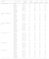
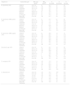
Of the antimicrobial agents tested, amikacin demonstrated the highest rate of susceptibility (%) for isolates of K. pneumoniae (92.2, 92.3), Enterobacter spp. (97.5, 92.1), and P. aeruginosa (85.3, 75.2) from both IAI (Table 1) and UTI (Table 2), respectively. Among K. pneumoniae, cephalosporins, piperacillin-tazobactam, and levofloxacin showed rates of susceptibility <67% for IAI isolates and <62% for UTI isolates. 11.8–22.8% of isolates were identified to be carbapenem-resistant. Jones et al. reported 9% of Klebsiella spp. as carbapenem-resistant in a regional surveillance study of 2011 data from Latin American patients with various infection types,7 suggesting dissemination of a successful clone or horizontal spread of resistance genes conferring carbapenem resistance. ESBL-positive isolates accounted for 42.0% of isolates of K. pneumoniae from IAI and 46.6% of isolates from UTI. Our result for isolates from IAI was similar to the rate reported from the SMART study for Latin America in 2008 (37.7%)8 but substantially higher than the rate reported from the same study in 2003 (14%).9 Ertapenem (68.5, 62.6) and imipenem (79.2, 75.9) demonstrated substantially higher rates of susceptibility (%) than the other β-lactams tested, including piperacillin-tazobactam (35.9, 37.4), against ESBL-positive isolates of K. pneumoniae from IAI and UTI, respectively. ESBL-positive K. pneumoniae were also less susceptible to amikacin and levofloxacin than were ESBL-negative isolates.
In vitro activity of antimicrobial agents against Gram-negative ESKAPE pathogens isolated from patients with intra-abdominal infections.
| Organism (n) | Antimicrobial agent | MIC range (μg/mL) | MIC90 (μg/mL) | % Susceptible | % Intermediate | % Resistant |
|---|---|---|---|---|---|---|
| K. pneumoniae (974) | Amikacin | ≤4 to >32 | 16 | 92.2 | 4.1 | 3.7 |
| Cefepime | ≤0.5 to >32 | >32 | 55.8 | 5.3 | 38.9 | |
| Ceftazidime | ≤0.5 to >32 | >32 | 57.4 | 3.6 | 39.0 | |
| Ceftriaxone | ≤1 to >32 | >32 | 54.2 | 0.3 | 45.5 | |
| Ertapenem | ≤0.03 to >4 | >4 | 81.8 | 1.3 | 16.8 | |
| Imipenem | ≤0.06 to >8 | 4 | 86.4 | 1.9 | 11.8 | |
| Levofloxacin | ≤0.5 to >4 | >4 | 66.9 | 2.5 | 30.6 | |
| Piperacillin-Tazobactam | ≤2 to >64 | >64 | 65.0 | 9.3 | 25.7 | |
| K. pneumoniae, ESBL-positive (409) | Amikacin | ≤4 to >32 | 32 | 87.0 | 7.1 | 5.9 |
| Cefepime | ≤0.5 to >32 | >32 | 6.1 | 11.7 | 82.2 | |
| Ceftazidime | ≤0.5 to >32 | >32 | 11.0 | 8.1 | 80.9 | |
| Ceftriaxone | ≤1 to >32 | >32 | 3.4 | 0.0 | 96.6 | |
| Ertapenem | ≤0.03 to >4 | >4 | 68.5 | 2.5 | 29.1 | |
| Imipenem | ≤0.06 to >8 | >8 | 79.2 | 2.9 | 17.9 | |
| Levofloxacin | ≤0.5 to >4 | >4 | 41.8 | 4.2 | 54.0 | |
| Piperacillin-Tazobactam | ≤2 to >64 | >64 | 35.9 | 17.6 | 46.5 | |
| K. pneumoniae, ESBL-negative (565) | Amikacin | ≤4 to >32 | ≤4 | 95.9 | 2.0 | 2.1 |
| Cefepime | ≤0.5 to >32 | 1 | 91.7 | 0.7 | 7.6 | |
| Ceftazidime | ≤0.5 to >32 | 1 | 91.0 | 0.4 | 8.7 | |
| Ceftriaxone | ≤1 to >32 | ≤1 | 91.0 | 0.5 | 8.5 | |
| Ertapenem | ≤0.03 to >4 | 0.06 | 91.5 | 0.5 | 8.0 | |
| Imipenem | ≤0.06 to >8 | 0.5 | 91.5 | 1.1 | 7.4 | |
| Levofloxacin | ≤0.5 to >4 | >4 | 85.1 | 1.2 | 13.6 | |
| Piperacillin-Tazobactam | ≤2 to >64 | >64 | 86.0 | 3.4 | 10.6 | |
| Enterobacter spp. (433) | Amikacin | ≤4 to >32 | ≤4 | 97.5 | 1.6 | 0.9 |
| Cefepime | ≤0.5 to >32 | >32 | 74.8 | 8.8 | 16.4 | |
| Ceftazidime | ≤0.5 to >32 | >32 | 65.1 | 3.2 | 31.6 | |
| Ceftriaxone | ≤1 to >32 | >32 | 59.6 | 2.1 | 38.3 | |
| Ertapenem | ≤0.03 to >4 | 1 | 83.6 | 6.7 | 9.7 | |
| Imipenem | 0.12 to >8 | 1 | 92.8 | 3.9 | 3.2 | |
| Levofloxacin | ≤0.5 to >4 | >4 | 87.1 | 2.8 | 10.2 | |
| Piperacillin-Tazobactam | ≤2 to >64 | >64 | 76.9 | 9.7 | 13.4 | |
| P. aeruginosa (570) | Amikacin | ≤4 to >32 | >32 | 85.3 | 1.2 | 13.5 |
| Cefepime | ≤0.5 to >32 | 32 | 77.4 | 9.1 | 13.5 | |
| Ceftazidime | ≤0.5 to >32 | 32 | 78.1 | 4.7 | 17.2 | |
| Imipenem | ≤0.06 to >8 | >8 | 72.3 | 2.8 | 24.9 | |
| Levofloxacin | ≤0.5 to >4 | >4 | 75.3 | 2.8 | 21.9 | |
| Piperacillin-Tazobactam | ≤2 to >64 | >64 | 77.7 | 11.2 | 11.1 | |
| A. baumannii (136) | Amikacin | ≤4 to >32 | >32 | 30.9 | 16.2 | 52.9 |
| Cefepime | ≤0.5 to >32 | >32 | 19.1 | 6.6 | 74.3 | |
| Ceftazidime | ≤0.5 to >32 | >32 | 21.3 | 2.9 | 75.7 | |
| Ceftriaxone | ≤1 to >32 | >32 | 11.0 | 11.0 | 77.9 | |
| Imipenem | 0.12 to >8 | >8 | 20.6 | 2.2 | 77.2 | |
| Levofloxacin | ≤0.5 to >4 | >4 | 15.4 | 7.4 | 77.2 | |
| Piperacillin-Tazobactam | ≤2 to >64 | >64 | 16.9 | 3.7 | 79.4 | |
In vitro activity of antimicrobial agents against Gram-negative ESKAPE pathogens isolated from patients with urinary tract infections.
| Organism (n) | Antimicrobial agent | MIC range (μg/mL) | MIC90 (μg/mL) | % Susceptible | % Intermediate | % Resistant |
|---|---|---|---|---|---|---|
| K. pneumoniae (597) | Amikacin | ≤4 to >32 | 16 | 92.3 | 3.7 | 4.0 |
| Cefepime | ≤0.5 to >32 | >32 | 49.6 | 4.2 | 46.2 | |
| Ceftazidime | ≤0.5 to >32 | >32 | 51.1 | 5.5 | 43.4 | |
| Ceftriaxone | ≤1 to >32 | >32 | 46.1 | 0.2 | 53.8 | |
| Ertapenem | ≤0.03 to >4 | >4 | 75.5 | 1.7 | 22.8 | |
| Imipenem | ≤0.06 to >8 | >8 | 81.4 | 2.4 | 16.3 | |
| Levofloxacin | ≤0.5 to >4 | >4 | 57.3 | 2.5 | 40.2 | |
| Piperacillin-Tazobactam | ≤2 to >64 | >64 | 61.5 | 9.6 | 29.0 | |
| K. pneumoniae, ESBL-positive (278) | Amikacin | ≤4 to >32 | 32 | 87.1 | 6.1 | 6.8 |
| Cefepime | ≤0.5 to >32 | >32 | 7.2 | 7.6 | 85.3 | |
| Ceftazidime | ≤0.5 to >32 | >32 | 11.2 | 10.1 | 78.8 | |
| Ceftriaxone | ≤1 to >32 | >32 | 1.8 | 0.4 | 97.8 | |
| Ertapenem | ≤0.03 to >4 | >4 | 62.6 | 3.6 | 33.8 | |
| Imipenem | ≤0.06 to >8 | >8 | 75.9 | 4.3 | 19.8 | |
| Levofloxacin | ≤0.5 to >4 | >4 | 34.5 | 4.0 | 61.5 | |
| Piperacillin-Tazobactam | ≤2 to >64 | >64 | 37.4 | 17.3 | 45.3 | |
| K. pneumoniae, ESBL-negative (319) | Amikacin | ≤4 to >32 | ≤4 | 96.9 | 1.6 | 1.6 |
| Cefepime | ≤0.5 to >32 | >32 | 86.5 | 1.3 | 12.2 | |
| Ceftazidime | ≤0.5 to >32 | >32 | 85.9 | 1.6 | 12.5 | |
| Ceftriaxone | ≤1 to >32 | >32 | 84.6 | 0.0 | 15.4 | |
| Ertapenem | ≤0.03 to >4 | >4 | 86.8 | 0.0 | 13.2 | |
| Imipenem | ≤0.06 to >8 | >8 | 86.2 | 0.6 | 13.2 | |
| Levofloxacin | ≤0.5 to >4 | >4 | 77.1 | 1.3 | 21.6 | |
| Piperacillin-Tazobactam | ≤2 to >64 | >64 | 82.5 | 2.8 | 14.7 | |
| Enterobacter spp. (165) | Amikacin | ≤4 to >32 | 16 | 92.1 | 3.0 | 4.9 |
| Cefepime | ≤0.5 to >32 | >32 | 68.5 | 3.6 | 27.9 | |
| Ceftazidime | ≤0.5 to >32 | >32 | 60.0 | 4.2 | 35.8 | |
| Ceftriaxone | ≤1 to >32 | >32 | 54.6 | 4.9 | 40.6 | |
| Ertapenem | ≤0.03 to >4 | 4 | 79.4 | 3.0 | 17.6 | |
| Imipenem | 0.12 to >8 | 2 | 86.1 | 6.7 | 7.3 | |
| Levofloxacin | ≤0.5 to >4 | >4 | 76.4 | 1.2 | 22.4 | |
| Piperacillin-Tazobactam | ≤2 to >64 | >64 | 72.1 | 8.5 | 19.4 | |
| P. aeruginosa (165) | Amikacin | ≤4 to >32 | >32 | 75.2 | 1.2 | 23.6 |
| Cefepime | ≤0.5 to >32 | >32 | 61.2 | 7.3 | 31.5 | |
| Ceftazidime | ≤0.5 to >32 | >32 | 62.4 | 7.3 | 30.3 | |
| Imipenem | 0.12 to >8 | >8 | 60.0 | 3.6 | 36.4 | |
| Levofloxacin | ≤0.5 to >4 | >4 | 55.2 | 6.1 | 38.8 | |
| Piperacillin-Tazobactam | ≤2 to >64 | >64 | 60.0 | 15.8 | 24.2 | |
| A. baumannii (43) | Amikacin | ≤4 to >32 | >32 | 16.3 | 25.6 | 58.1 |
| Cefepime | ≤1 to >32 | >32 | 11.6 | 9.3 | 79.1 | |
| Ceftazidime | 2 to >32 | >32 | 18.6 | 0.0 | 81.4 | |
| Ceftriaxone | 8 to >32 | >32 | 11.6 | 7.0 | 81.4 | |
| Imipenem | 0.12 to >8 | >8 | 18.6 | 0.0 | 81.4 | |
| Levofloxacin | ≤0.5 to >4 | >4 | 11.6 | 7.0 | 81.4 | |
| Piperacillin-Tazobactam | ≤2 to >64 | >64 | 18.6 | 2.3 | 79.1 | |
For Enterobacter spp. from IAI, imipenem, ertapenem, and levofloxacin demonstrated rates of susceptibility ranging from 83.6% to 92.8%, up to 33 percentage points higher than the susceptibility rates for cefepime, ceftazidime, ceftriaxone, and piperacillin-tazobactam (59.6–76.9%). A similar pattern was found for Enterobacter spp. from UTI. On the other hand, isolates of P. aeruginosa showed rates of susceptibility that were more uniform across cefepime, ceftazidime, imipenem, levofloxacin, and piperacillin-tazobactam (between 72.3% and 78.1% in isolates from IAI and between 55.2% and 62.4% in UTI isolates). The percentage of isolates of P. aeruginosa from IAI that were susceptible to amikacin, imipenem, cefepime, ceftazidime, piperacillin-tazobactam, and levofloxacin were similar to those reported from the SMART study for Latin America in 2008.8
Rates of susceptibility to all agents tested against A. baumannii from both infection sources were ≤30.9% (Tables 1 and 2). A. baumannii is an increasingly frequent pathogen in hospitalized patients with IAI and UTI.10 It is often multidrug-resistant, associated with patients in ICUs, demonstrates variability in its geographic prevalence, and frequently presents clinicians with therapeutic dilemmas given its resistance profile.10 In the current study as well as in another study describing A. baumannii from other regions of the world amikacin has been shown to be the most active antimicrobial agent against A. baumannii among the agents tested in the SMART program.10
The proportion of Gram-negative ESKAPE pathogens among all Gram-negative isolates collected from IAI samples was 42.9% (564 isolates of the ESKAPE group among 1314 Gram-negative isolates) for isolates from patients located in an ICU compared with only 34.8% (1549/4450) for isolates from patients in non-ICU wards (data not shown). The difference observed in the proportion of Gram-negative ESKAPE pathogens among all Gram-negative isolates was even greater for isolates from UTI where 47.8% (274/573) of isolates from ICUs were Gram-negative ESKAPE pathogens versus 32.8% (696/2125) of isolates from non-ICU locations (data not shown). These results are consistent with the finding of another study that the majority of pathogens isolated from ICUs in some Latin American countries are from the ESKAPE group.11
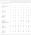
An analysis comparing rates of antimicrobial susceptibility for isolates of Gram-negative ESKAPE pathogens grouped into those isolated from patients in ICUs and those isolated from hospitalized patients outside of ICUs (non-ICU) was also performed (Table 3). ESBL-positive rates of K. pneumoniae were higher in isolates from ICU patients for both IAI (44.7%) and UTI (52.9%) than for isolates from patients in non-ICU settings with IAI (41.0%) and UTI (44.4%). In general, rates of susceptibility to the antimicrobial agents tested were lower for isolates from the ICU than for those isolated from hospitalized patients outside of the ICU, with a few exceptions such as, for example, levofloxacin against Enterobacter spp. from both IAI and UTI and all antimicrobial agents against P. aeruginosa from UTI. Higher resistance and ESBL-positive rates in ICUs than non-ICU wards are consistent with findings in other studies and some other global regions (for example in Europe),12–14 but not necessarily in all regions (for example, ICU/non-ICU differences were not found to be as marked in a recent study of SMART isolates from Asia/Pacific).15
In vitro activity of antimicrobial agents against Gram-negative ESKAPE pathogens isolated from patients in ICUs and non-ICU wards.
| Source/organism/patient location (n) | % Susceptible | |||||||
|---|---|---|---|---|---|---|---|---|
| AMK | FEP | CAZ | CRO | ETP | IPM | LVX | TZP | |
| Intra-abdominal infections | ||||||||
| K. pneumonia | ||||||||
| ICU (262) | 90.1 | 49.6 | 53.1 | 50.0 | 76.7 | 80.5 | 65.3 | 56.9 |
| Non-ICU (712) | 93.0 | 58.0 | 59.0 | 55.8 | 83.7 | 88.5 | 67.6 | 68.0 |
| K. pneumoniae, ESBL-positive | ||||||||
| ICU (117) | 84.6 | 4.3 | 12.0 | 4.3 | 63.3 | 71.8 | 43.6 | 29.9 |
| Non-ICU (292) | 88.0 | 6.9 | 10.6 | 3.1 | 70.6 | 82.2 | 41.1 | 38.4 |
| K. pneumoniae, ESBL-negative | ||||||||
| ICU (145) | 94.5 | 86.2 | 86.2 | 86.9 | 87.6 | 87.6 | 82.8 | 78.6 |
| Non-ICU (420) | 96.4 | 93.6 | 92.6 | 92.4 | 92.9 | 92.9 | 86.0 | 88.6 |
| Enterobacter spp. | ||||||||
| ICU (112) | 96.4 | 69.6 | 53.6 | 48.2 | 72.3 | 92.0 | 87.5 | 67.0 |
| Non-ICU (321) | 97.8 | 76.6 | 69.2 | 63.6 | 87.5 | 93.2 | 86.9 | 80.4 |
| P. aeruginosa | ||||||||
| ICU (142) | 81.0 | 71.1 | 73.9 | NA | NA | 62.7 | 72.5 | 69.7 |
| Non-ICU (428) | 86.7 | 79.4 | 79.4 | NA | NA | 75.5 | 76.2 | 80.4 |
| A. baumannii | ||||||||
| ICU (48) | 33.3 | 14.6 | 16.7 | 6.3 | NA | 14.6 | 8.3 | 10.4 |
| Non-ICU (88) | 29.6 | 21.6 | 23.9 | 13.6 | NA | 23.9 | 19.3 | 20.5 |
| Urinary tract infections | ||||||||
| K. pneumonia | ||||||||
| ICU (153) | 91.5 | 41.2 | 43.1 | 38.6 | 70.6 | 76.5 | 52.9 | 53.6 |
| Non-ICU (444) | 92.6 | 52.5 | 53.8 | 48.7 | 77.3 | 83.1 | 58.8 | 64.2 |
| K. pneumoniae, ESBL-positive | ||||||||
| ICU (81) | 87.7 | 4.9 | 8.6 | 1.2 | 60.5 | 72.8 | 33.3 | 34.6 |
| Non-ICU (197) | 86.8 | 8.1 | 12.2 | 2.0 | 63.5 | 77.2 | 35.0 | 38.6 |
| K. pneumoniae, ESBL-negative | ||||||||
| ICU (72) | 95.8 | 81.9 | 81.9 | 80.6 | 81.9 | 80.6 | 75.0 | 75.0 |
| Non-ICU (247) | 97.2 | 87.9 | 87.0 | 85.8 | 88.3 | 87.9 | 77.7 | 84.6 |
| Enterobacter spp. | ||||||||
| ICU (49) | 95.9 | 65.3 | 53.1 | 49.0 | 75.5 | 89.8 | 81.6 | 65.3 |
| Non-ICU (116) | 90.5 | 69.8 | 62.9 | 56.9 | 81.0 | 84.5 | 74.1 | 75.0 |
| P. aeruginosa | ||||||||
| ICU (54) | 79.6 | 70.4 | 64.8 | NA | NA | 66.7 | 66.7 | 70.4 |
| Non-ICU (111) | 73.0 | 56.8 | 61.3 | NA | NA | 56.8 | 49.6 | 55.0 |
| A. baumannii | ||||||||
| ICU (18) | 22.2 | 5.6 | 16.7 | 5.6 | NA | 16.7 | 5.6 | 16.7 |
| Non-ICU (25) | 12.0 | 16.0 | 20.0 | 16.0 | NA | 20.0 | 16.0 | 20.0 |
AMK, amikacin; FEP, cefepime; CAZ, ceftazidime; CRO, ceftriaxone; ETP, ertapenem; IPM, imipenem; LVX, levofloxacin; TZP, piperacillin-tazobactam; NA, not applicable (CLSI breakpoints are not available for ceftriaxone and ertapenem tested against P. aeruginosa and for ertapenem tested against A. baumannii).
An analysis of antimicrobial resistance by country was difficult because of low number of participating sites and small sample sizes of collected isolates per country. We were able to at least examine the ESBL-positive rate of K. pneumoniae when both infection sources were combined for countries with two or more sites and at least 100 isolates. Rates ranged from 31.2% for isolates collected from sites in Colombia (48/154), 34.7% in Venezuela (61/176), 36.1% in Panama (43/119), 38.4% in Mexico (101/263), and 44.6% in Argentina (70/157) to 50.5% in Brazil (142/281), and 69.3% in Chile (104/150). Jones et al. found a similarly substantial variation across countries in ESBL-positive rates among Klebsiella spp. in the Latin America surveillance study from 2011.7 When comparing the countries that participated in both the 2011 study and the current study and again examining only countries with at least two sites and 100 isolates, Venezuela (40% ESBL-positive Klebsiella spp. in 2011) was among the countries with the lowest rates in both studies, while Chile (59% in 2011) demonstrated the highest rate in both studies.7
The SMART global surveillance program has monitored the in vitro antimicrobial susceptibility profiles of clinical isolates of Gram-negative bacilli collected worldwide from patients with IAI since 2002 and from patients with UTI since late 2009 against a uniform list of parenteral antimicrobial agents.4 The antimicrobial agents tested in the SMART global surveillance program are those recommended by the Surgical Infection Society and the Infectious Diseases Society of America in their guidelines for the diagnosis and management of complicated IAI16 and may also potentially be prescribed for hospitalized patients with UTI attributable to Gram-negative ESKAPE pathogens.
Previously published summaries of surveillance studies have noted that Latin American countries, in general, tend to demonstrate higher rates of antimicrobial resistance for many key bacterial pathogens, including ESKAPE pathogens, compared with European countries and the United States.17 The current study also found that Gram-negative ESKAPE pathogens isolated from Latin America demonstrated compromised in vitro susceptibility to commonly prescribed broad-spectrum, parenteral antimicrobial agents. The publication of such surveillance study data describing regional antimicrobial susceptibility/resistance rates in clinical isolates of Gram-negative ESKAPE pathogens is essential to stimulate antimicrobial stewardship efforts as well as to identify emerging resistance trends and geographic diversity over time.
Limitations of the current study include that the SMART surveillance program collects isolates from only a small number of sites per country and that the sites included are not uniformly distributed in all countries. In addition, the number of isolates collected at each site may vary over time and sites may not participate each year of the study. It is also important to note that isolate susceptibilities are defined in terms of MICs, and in vitro susceptibility does not necessarily translate into a successful clinical outcome. Furthermore, classification of isolates by patient location in the hospital is imperfect as patients frequently move between wards during a single admission.
The slowness to market of novel antimicrobial agents with reliable activity against Gram-negative ESKAPE pathogens suggests efforts to identify optimal strategies for infection control and prevention as well as antimicrobial use/stewardship need to intensify, especially in ICUs.3 Ongoing surveillance data is crucial as it provides guidance for empiric antimicrobial agent selection by identifying the most common pathogens and their antimicrobial susceptibility profiles. In addition to ongoing surveillance efforts, data on the impact of clinical interventions to decrease the prevalence of resistance are required. As resistance to parenteral broad-spectrum antimicrobial agents continues to increase, combination empiric therapies for infections potentially attributable to Gram-negative ESKAPE pathogens may become routine and will be driven by surveillance initatives.7
FundingFunding for this research was provided by Merck & Co., Inc., Kenilworth, NJ, which also included compensation fees for services in relation to preparing the manuscript.
Conflicts of interestJAK is an employee of the University of Manitoba and Diagnostic Services Manitoba and a consultant for International Health Management Associates, Inc. (IHMA, Inc.), which receives the funding for the SMART surveillance program. DJH is an employee of IHMA, Inc. and of the University of Manitoba and Diagnostic Services. MAH, SHL, and DFS are employees of IHMA, Inc. JAK and the IHMA authors do not have personal financial interests in the sponsor of this paper (Merck & Co., Inc.).
The authors thank all SMART participants in Latin America for their contributions to the program. Isolates from the Latin American region were submitted to the SMART program by clinical laboratories in Argentina, Brazil, Chile, Columbia, Dominican Republic, Ecuador, Guatemala, Mexico, Panama, Puerto Rico, and Venezuela.
