
We report an outbreak of Achromobacter xylosoxidans at a neonatal intensive care unit. We aimed to present clinical, laboratory and treatment data of the patients.
Materials and methodsAll consecutive episodes of bacteremia due to A. xylosoxidans at our neonatal intensive care unit, beginning with the index case detected at November 2009 until cessation of the outbreak in April 2010, were evaluated retrospectively.
ResultsThirty-four episodes of bacteremia occurred in 22 neonates during a 6-month period. Among the affected, 90% were preterm newborns with gestational age of 32 weeks or less and 60% had birth weight of 1000g or less. Endotracheal intubation, intravenous catheter use, total parenteral nutrition and prolonged antibiotic therapy were the predisposing conditions. Presenting features were abdominal distention, thrombocytopenia and neutropenia. The mortality rate was 13.6% and the majority of isolates were susceptible to piperacillin-tazobactam, carbapenems and trimethoprim-sulfametoxazole, and resistant to gentamycin. More than half were breakthrough infections. Despite intensive efforts to control the outbreak by standard methods of hand hygiene, patient screening and isolation, containment could be achieved only after the neonatal intensive care unit was relocated. The investigation was not able to single out the source of the outbreak.
ConclusionA. xylosoxidans has the potential to cause serious infections in premature babies. More studies are needed to determine the importance of different sources of infection in hospital units.
Achromobacter xylosoxidans is a nonfermentative Gram-negative bacillus which is widely distributed in nature, especially in aquatic environments.1,2 It was first described by Yabuuchi and Ohyama in 1971 from purulent ear discharge of patients with chronic otitis media.3 Since it is an opportunistic pathogen it usually affects patients with underlying illnesses and immunosuppression.4,5 Infections such as bacteremia, pneumonia, meningitis, urinary tract infections, abscesses, osteomyelitis, corneal ulcers, prosthetic valve endocarditis and peritonitis have been reported.1,4–11 Treatment of Achromobacter infections is difficult since the bacterium may be resistant to several antibiotics.2
Because of limited data and lack of large series of infections caused by Achromobacter in children, especially in newborns, we aimed to present an outbreak of A. xylosoxidans bacteremia that took place at our neonatal intensive care unit (NICU).
MethodsThe study was performed in a level III neonatal reference unit of a tertiary care hospital where a mean of 400 newborns are admitted annually. All consecutive episodes of bacteremia due to A. xylosoxidans, beginning with the index case detected in November 2009 until cessation of the outbreak in April 2010 were evaluated retrospectively. A case of A. xylosoxidans bacteremia was defined as one or more blood cultures positive for A. xylosoxidans when clinical signs of sepsis were present.12 Blood samples were incubated into Bactec 9240 system (Becton Dickinson, USA) for up to seven days. A sample from the blood culture bottle with positive bacterial growth was inoculated into 5% sheep blood agar and EMB (eosin methylene blue) agar (Oxoid, England), and incubated at 36.5°C for 18–20h. Gram negative bacilli with positive reaction for oxidase were detected in Gram-stained smears prepared from colonies. Identification of the microorganisms was done by API 20 NE system (bioMerieux, France). Susceptibility of the organisms to antimicrobial agents was performed by using (Kirby–Bauer) disk-diffusion method on Mueller–Hinton agar, according to the Clinical Laboratory Standards Institute (CLSI) Guidelines. Multiple episodes were indicated by the finding of A. xylosoxidans isolates in blood cultures obtained >four weeks apart, or >two weeks apart if the blood cultures became sterile and/or there was evidence of clinical resolution of the infection.4
Data recorded included sex, gestational age, birth weight, date of admission, underlying diseases, presence of intravascular catheters, administration of broad-spectrum antibiotics and total parenteral nutrition, number of positive blood cultures and sites of other cultures from which A. xylosoxidans was isolated. Neutropenia was defined as a count less than 1.5×10⁹/Lcells/mm3, thrombocytopenia was defined as a platelet count of <80×109/L.13 Infections that occurred more than 72h after admission were defined as nosocomial.14 A breakthrough infection was indicated by the isolation of bacteria from blood cultures obtained while the patient was receiving systemic antibiotics to which the bacteria was later confirmed to be susceptible.4
For the investigation of outbreak, bacteriological cultures of environmental samples were taken including antiseptic solutions (chlorhexidine digluconate, aqueous and alcoholic), wash basins, incubator surfaces, liquid soaps, faucets, nebulizators and tap water. Hands of health care workers were sampled by direct fingerprinting in Petri dishes. Fluids were filtered through 0.45μm membranes (Millipore, Bedford, MA, USA) and cultivated on sheep blood agar (SLB, Turkey), EMB agar. Swabs were plated on EMB agar and 5% sheep blood agar. Plates were incubated for 24h at 36°C. Environmental cultures were taken following detection of first four bacteremia cases in November 2009 and discontinued after April 2010.
ResultsIndex caseThe index case was a 25-week gestational age male (birth weight 660g) baby born to a G1P0A0 mother. After birth he was intubated and transferred to the NICU with a diagnosis of respiratory distress syndrome (RDS). He received ampicillin-sulbactam and gentamycin since congenital pneumonia could not be excluded. On the 7th day of life, minimal enteral feeding was initiated but in the next day abdominal distension developed and antibiotics were switched to vancomycin and meropenem with a diagnosis of necrotizing enterocolitis (NEC). Blood cultures remained sterile and vancomycin was discontinued after 14 days. On 21st day of meropenem therapy, he developed apnea, leukopenia and thrombocytopenia. He was reintubated and meropenem was switched to sulbactam-cefoperazone and fluconazole pending culture results. His clinical condition deteriorated and he developed acidosis, hypotension and bradycardia necessitating a short duration of adrenalin infusion. Two sets of blood cultures yielded A. xylosoxidans susceptible to ciprofloxacin, meropenem and trimethoprim-sulfametoxazole (TMP-SXM). Sulbactam-cefoperazone and fluconazole were discontinued and meropenem plus ciprofloxacin were given. Blood cultures became sterile after five days. The patient's clinical condition resolved gradually and he was extubated after seven days. He was discharged from the NICU when at the age of two months.
Epidemiologic investigationFollowing the index case, four additional cases were diagnosed within the same week. An outbreak was suspected and cohorting and contact precautions with gown and glove use on entry to rooms of patients infected with A. xylosoxidans and enhanced environmental cleaning were instituted. An investigation for determination of source of infection was started. Standard infection control practices such as hand hygiene (hand washing upon unit entry before and after patient contact and use of alcohol hand sanitizer between patient contacts), aseptic technique for all invasive procedures, sterile procedures requiring gown, mask, and gloves and environmental cleaning were overviewed. Education of healthcare staff was repeated but no flaw was found. Environmental vectors were suspected and bacteriological cultures of environmental samples were taken but the source could not be identified. Twelve patients were determined to be infected during November and December. A second attack affecting 10 patients was observed in March. Patients were transferred to an other part of the hospital prepared as NICU in April. No further cases were detected during the following 12 months.
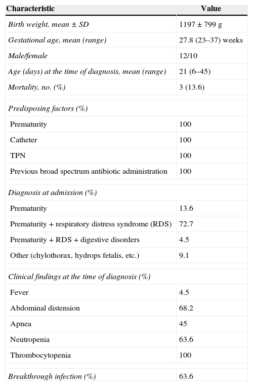
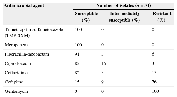
Clinical features and treatment of affected neonatesA total of 22 neonates, 12 (54.5%) male, were diagnosed with A. xylosoxidans bacteremia. All infecting strains were considered nosocomially acquired. All patients were preterm and 60% were under 1000g birth weight. Prematurity and respiratory distress syndrome (RDS) were the most common diagnosis at admission. Most common presenting features of infection were abdominal distention, apnea, thrombocytopenia and leukopenia (Table 1). Three out of 22 patients had cerebrospinal fluid (CSF) abnormalities suggestive of bacterial meningitis and A. xylosoxidans growth was detected in CSF in addition to blood culture. 84% of patients recovered with antibiotic therapy while three patients died due to Achromobacter sepsis. Two patients had multiple growths of A. xylosoxidans. In vitro susceptibility profile of A. xylosoxidans isolates to antimicrobial agents is given in Table 2. Breakthrough infection was observed in 14 patients (63.6%). Patients received 10–21 day course of meropenem alone (9.7%), meropenem in combination with other antibiotics (meropenem plus ciprofloxacin, meropenem plus ceftazidime and meropenem plus piperacillin-tazobactam in 50%, 22.7%, and 13.6% of patients, respectively) or ciprofloxacin plus ceftazidime (4.5%).
Characteristics of 22 newborns infected with Achromobacter xylosoxidans.
| Characteristic | Value |
|---|---|
| Birth weight, mean±SD | 1197±799g |
| Gestational age, mean (range) | 27.8 (23–37) weeks |
| Male/female | 12/10 |
| Age (days) at the time of diagnosis, mean (range) | 21 (6–45) |
| Mortality, no. (%) | 3 (13.6) |
| Predisposing factors (%) | |
| Prematurity | 100 |
| Catheter | 100 |
| TPN | 100 |
| Previous broad spectrum antibiotic administration | 100 |
| Diagnosis at admission (%) | |
| Prematurity | 13.6 |
| Prematurity+respiratory distress syndrome (RDS) | 72.7 |
| Prematurity+RDS+digestive disorders | 4.5 |
| Other (chylothorax, hydrops fetalis, etc.) | 9.1 |
| Clinical findings at the time of diagnosis (%) | |
| Fever | 4.5 |
| Abdominal distension | 68.2 |
| Apnea | 45 |
| Neutropenia | 63.6 |
| Thrombocytopenia | 100 |
| Breakthrough infection (%) | 63.6 |
In vitro susceptibility profile of 34 Achromobacter xylosoxidans isolates to antimicrobial agents.
| Antimicrobial agent | Number of isolates (n=34) | ||
|---|---|---|---|
| Susceptible (%) | Intermediately susceptible (%) | Resistant (%) | |
| Trimethoprim-sulfametoxazole (TMP-SXM) | 100 | 0 | 0 |
| Meropenem | 100 | 0 | 0 |
| Piperacillin-tazobactam | 91 | 3 | 6 |
| Ciprofloxacin | 82 | 15 | 3 |
| Ceftazidime | 82 | 3 | 15 |
| Cefepime | 15 | 9 | 76 |
| Gentamycin | 0 | 0 | 100 |
A. xylosoxidans is an emerging nosocomial pathogen. It represented 12.5% of nonfermenting Gram negative bacilli isolated in Latin America medical centers during SENTRY program 1997–2002.15 Respiratory tract colonization in cystic fibrosis patients has also been recognized during the last decade.16 Case reports due to A. xylosoxidans infection in Turkey are increasingly reported.17–19 Nevertheless, knowledge about neonatal infections caused by A. xylosoxidans is relatively scarce. It was reported as a contaminant leading to pseudobacteremia outbreak at NICU but serious infections such as meningitis and fatal sepsis in prematures were also described.6,20–22
A. xylosoxidans bacteremia is almost always a nosocomial infection.23 Bacteremia due to A. xylosoxidans is usually related to intravascular catheters and is frequently polymicrobial in patients with underlying malignancies.4,24 Shie et al. evaluated characteristics of A. xylosoxidans bacteremia in Taiwan and reported that malignancies, central venous catheter implants and previous major surgery were the most common underlying conditions.25 Another study in a tertiary care hospital in India identified 20 strains of A. xylosoxidans isolated from 15 patients during one year period.26 Seventeen isolates were from blood, two from CSF and one from pleural fluid; 30% of patients were premature neonates. We identified low birth weight, prematurity, total parenteral nutrition (TPN) and previous broad spectrum antibiotic therapy as the major predisposing conditions for Achromobacter bacteremia in our study. 4.5% of patients had undergone surgical interventions. The mean interval from hospital admission to isolation of a positive culture was 21 days. All affected neonates had umbilical catheter insertion and in all bacteremia episodes A. xylosoxidans was isolated as the sole pathogen.
Aisenberg et al. reported that Achromobacter bacteremia presented with fever (52%), sepsis syndrome (22%) and respiratory complaints (17%).4 In another report of 40 cases of A. xylosoxidans bacteremia, the most common manifestations were fever (82%), chills (37.5%), hypotension (32.5%) and altered consciousness.25 Dyspnea, abdominal pain, oliguria and diarrhea were also noted. In our study, thrombocytopenia (100%), neutropenia (63.6), abdominal distension (69%) and apnea (45%) were the most common clinical features. These findings are common with other Gram negative bacilli and fungi septicemia which makes the diagnosis rather difficult unless proven by culture. Fever was reported less frequently (4.5%) probably because of insufficient immune response of premature neonates. Three deaths were attributable to A. xylosoxidans sepsis with a fatality rate lower than previous reports (15–47.5%).4,23,25
There have been many reports of A. xylosoxidans isolation from environmental sources in hospital acquired infections.23A. xylosoxidans was detected in deionized water in an intensive care unit and tap water and on the hands of two health care workers in a hemodialysis unit.1,23 We checked environmental samples and hands of healthcare personnel for colonization but were unable to determine the source. In a similar report from India, Kumar et al. were also unable to determine the source.26 Since there was similarity of antibiotic susceptibility pattern of isolates, probable clonality of the isolates could be predicted but we were unable to perform testing for molecular epidemiology. In addition to enhanced infection control measures and increased awareness of health care staff, decreased patient capacity during the first days following relocation of NICU may have prevented development of additional cases of A. xylosoxidans bacteremia.
A. xylosoxidans has an unusual susceptibility pattern with resistance to aminoglycosides and to the majority of β-lactam antibiotics. Anti-pseudomonal penicillins, TMP-SMZ and carbapenems are the preferred agents in treatment.2,27 All of our isolates were sensitive to TMP-SXM and meropenem and the majority were sensitive to piperacillin-tazobactam similar to previous reports. Susceptibility to the fluoroquinolones is variable.28 Gomez et al. and Almuzara et al. have reported that quinolones have poor activity against Achromobacter spp.23,29 In contrast 82% of our isolates were susceptible to ciprofloxacin. For infections caused by multidrug-resistant A. xylosoxidans, antimicrobial combinations such as piperacillin plus gentamycin, azitromycin plus doxycycline, and azitromycin plus TMP-SXM have been tested with favorable results.24 Since 63% of infections were definite breakthrough infections and the patients were severely ill we used combination therapy in most patients.
ConclusionA. xylosoxidans has the potential to cause serious infections in hospitalized neonates. Antimicrobial susceptibility profile for each case should be taken into account for determining the therapy. More studies are needed to assess the importance of different sources of contamination in hospital units.
Conflict of interestThe authors declare no conflicts of interest.
