Journal Information
Vol. 27. Issue 3.
(May - June 2023)
Share
Share
Download PDF
More article options
Vol. 27. Issue 3.
(May - June 2023)
Clinical Image
Full text access
Tapeworm (Diphyllobothrium nihonkaiense) detected by abdominal ultrasonography
Visits
819
Ryota Hasea,b,
Corresponding author
hase.ryota@kameda.jp

Corresponding author.
, Haruki Mitoa, Yudai Yanoa, Yasuyuki Morishimac, Yuichi Hasegawad
a Japanese Red Cross Narita Hospital, Department of Infectious Diseases, Narita, Chiba, Japan
b Kameda Medical Center, Department of Infectious Diseases, Kamogawa, Chiba, Japan
c National Institute of Infectious Diseases, Department of Parasitology, Tokyo, Japan
d Japanese Red Cross Narita Hospital, Department of Laboratory Medicine, Narita, Chiba, Japan
This item has received
Article information
Full Text
Bibliography
Download PDF
Statistics
Figures (2)
Full Text

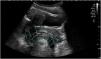
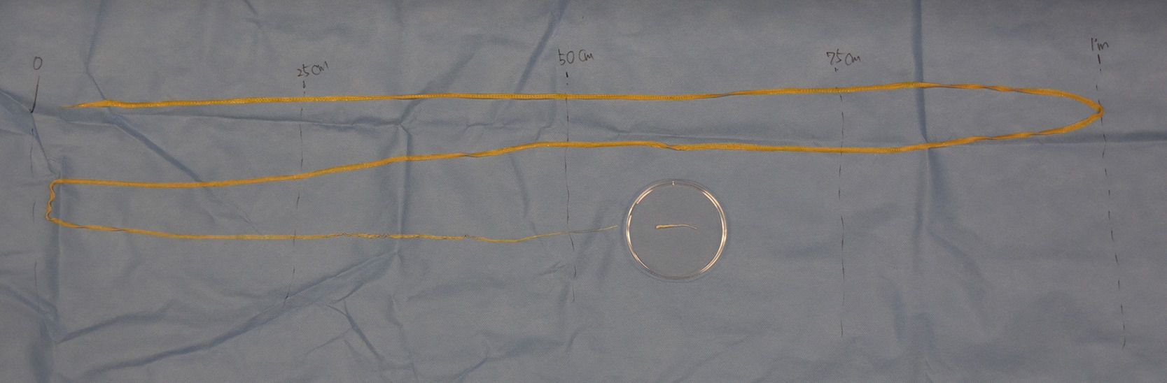
A 16-year-old girl presented with anal discharge of a white thread-like object. She denied additional symptoms. She had consumed raw salmon three weeks ago. Physical examination revealed no abnormal findings, and blood tests were unremarkable. However, abdominal ultrasound revealed a hyperechoic strand-like structure (Fig. 1). Stool examination revealed the presence of cestode ova. Therefore, she was admitted to our hospital and treated with oral praziquantel. A tapeworm, roughly 2.5 m long, was extracted from her feces (Fig. 2). Restriction fragment length polymorphism analysis with PCR-amplified cox1 gene fragment was performed,1 which identified the causative cestode was Diphyllobothrium nihonkaiense.

Fig. 1.

Longitudinal view of the rectum showing high echoic strand-like lesion (blue arrows). (For interpretation of the references to color in this figure legend, the reader is referred to the web version of this article.)

(0.16MB).
Fig. 2.

The tapeworm unfolded after deworming with praziquantel.

(0.08MB).

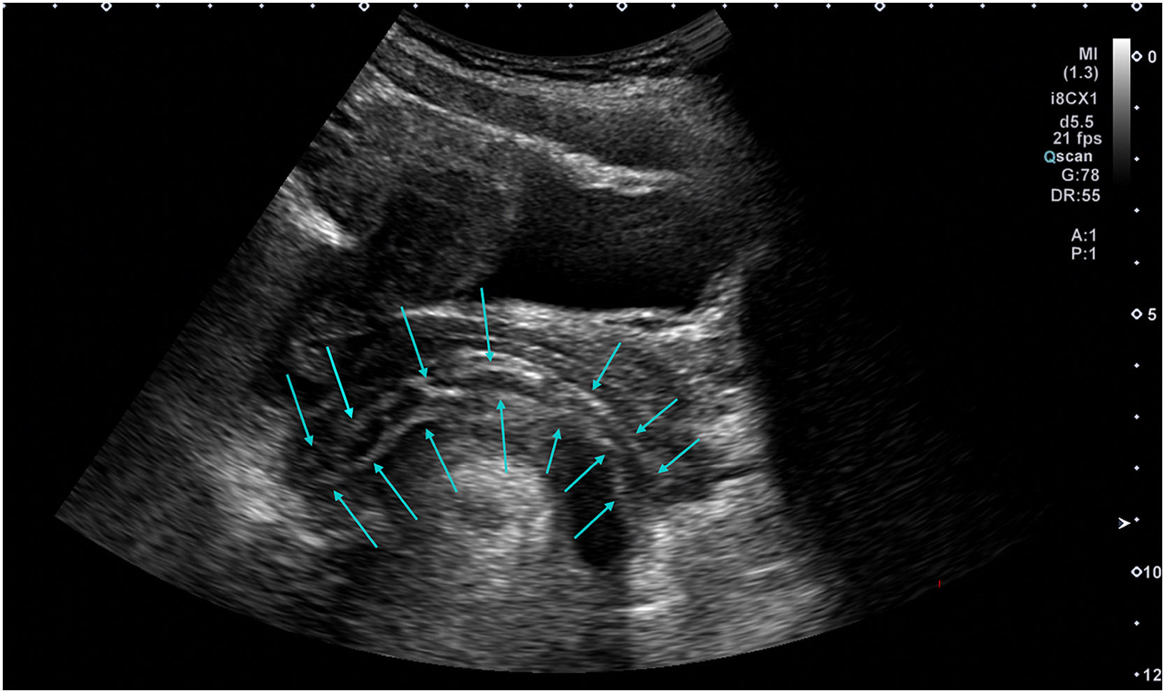
In Japan, D. nihonkaiense is the most common diphyllobothriasis-causing tapeworm.2 The diagnosis was based on the appearance after deworming, and the species was correctly identified using a molecular technique. The effectiveness of ultrasound has been reported in several publications.3,4 In cases such ours, ultrasound is useful for deciding whether to treat patients with praziquantel because the tapeworms are extracted naturally. In conclusion, abdominal ultrasonography can be useful for diagnosing diphyllobothriasis.

References
[1]
H. Yamasaki, D. Tsubokawa, R. Mercado, T. Kuramochi.
A simple method for identifying the diphyllobothriids based on mitochondrial DNA analysis.
Materials and Methods in Parasitology, pp. 47-49
[2]
H. Ikuno, S. Akao, H. Yamasaki.
Epidemiology of Diphyllobothrium nihonkaiense diphyllobothriasis, Japan, 2001-2016.
Emerg Infect Dis, 24 (2018), pp. 1428-1434
[3]
M. Hiroshima, M. Nakagawa, Y. Ito, A. Shiba, Y. Ozawa, Y. Shibamoto.
Ultrasonographic findings of Diphyllobothrium nihonkaiense: a case report.
Radiol Case Rep, 14 (2018), pp. 63-65
[4]
H. Kitamoto, S. Inoue, K. Okamoto, T. Inokuma.
Scanning early catches the worm: abdominal ultrasound as a possible screening method for intestinal cestodes.
Copyright © 2023. Sociedade Brasileira de Infectologia
The Brazilian Journal of Infectious Diseases
Article options
Tools